3.10.6.1 - Imaging Techniques
A radioactive tracer makes the distribution or behaviour of a substance visible. Learn how tracer chemistry, nuclear properties and detector geometry work together, from a hospital technetium generator to PET image reconstruction.
Gamma-emitting radioisotopes as tracers
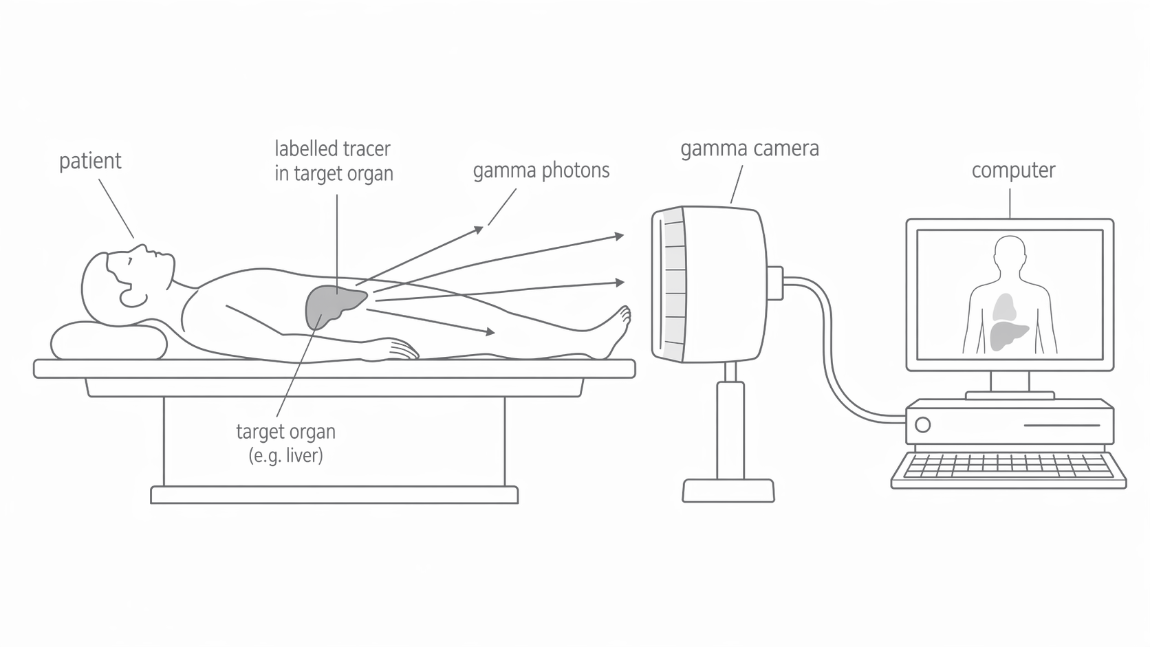
A radioactive tracer follows a biological process because its chemical form has an affinity for the tissue being investigated. A radionuclide can label a compound or blood cells; iodine itself is taken up by the thyroid. After administration, some emitted gamma photons escape the body and are detected externally. Their distribution reveals function, such as uptake or blood flow, rather than just the outline of an organ.
Gamma radiation is suitable for external detection because it is penetrating. It is still ionising: photons absorbed or scattered in tissue contribute to dose. Alpha and beta particles deposit energy over shorter distances and usually cannot provide the signal for an external gamma camera. A tracer therefore needs a useful gamma signal without unnecessary locally absorbed radiation.
[DIAGRAM: asset_name: B.6.1 - Imaging Techniques - Diagram 1; asset_slug: B.6.1 - Imaging Techniques - Diagram 1; caption: Gamma photons from an internal tracer reach an external detector; description: Gamma photons from an internal tracer reach an external detector.]
Matching the isotope to the investigation
The half-life must allow preparation, uptake and imaging, while avoiding unnecessarily prolonged activity. Photon energy must permit escape from tissue and efficient detection; very high energies are harder to absorb and collimate. Chemistry must carry the radionuclide to the relevant organ. A good half-life cannot compensate for a tracer that does not reach the target.
| Radionuclide | Main radiation relevant here | Physical half-life | Principal gamma energies | Targeting example |
|---|---|---|---|---|
| Technetium-99m | Gamma emission from an excited nuclear state | About | Many labelled compounds, including those used for bone, kidney and heart imaging | |
| Iodine-131 | Beta-minus and gamma | About | About , often rounded to | Iodine is taken up by the thyroid |
| Indium-111 | Electron capture followed by gamma emission | About | About and , often rounded to and | Labelled white blood cells or antibodies for slower investigations |
The m in technetium-99m means metastable, an excited nuclear state. Technetium-99m and indium-111 provide gamma radiation without a beta-minus spectrum, an advantage for imaging. The school shorthand “gamma emitter” does not mean zero dose or literally no other emissions: internal-conversion electrons, atomic X-rays and Auger electrons can occur. Detailed branching fractions are not needed here.
Iodine-131's thyroid uptake is useful, but its beta emission adds local tissue dose. It is important in thyroid treatment and some imaging; it is not automatically the preferred isotope for every diagnostic thyroid scan. Indium's longer half-life suits slower cell tracking, while technetium's range of labelling chemistry suits many same-day investigations.
The molybdenum-technetium generator
A molybdenum-technetium generator transports a longer-lived parent to the hospital and provides a short-lived daughter there. Molybdenum-99 has a physical half-life of about , compared with for technetium-99m. The parent therefore remains useful during delivery and subsequent production, although it too decays and the generator eventually needs replacement.
Molybdenum-99 is bound to an aluminium oxide column inside shielding. Its beta-minus decay supplies technetium-99m. The technetium is less strongly bound and is washed from the column with sterile saline: this separation is elution. Most molybdenum remains on the column and continues producing daughter nuclei. The collected technetium can then be prepared in a suitable radiopharmaceutical.
[DIAGRAM: asset_name: B.6.1 - Imaging Techniques - Diagram 2; asset_slug: B.6.1 - Imaging Techniques - Diagram 2; caption: Saline elutes technetium while molybdenum stays on the shielded column; description: Saline elutes technetium while molybdenum stays on the shielded column.]
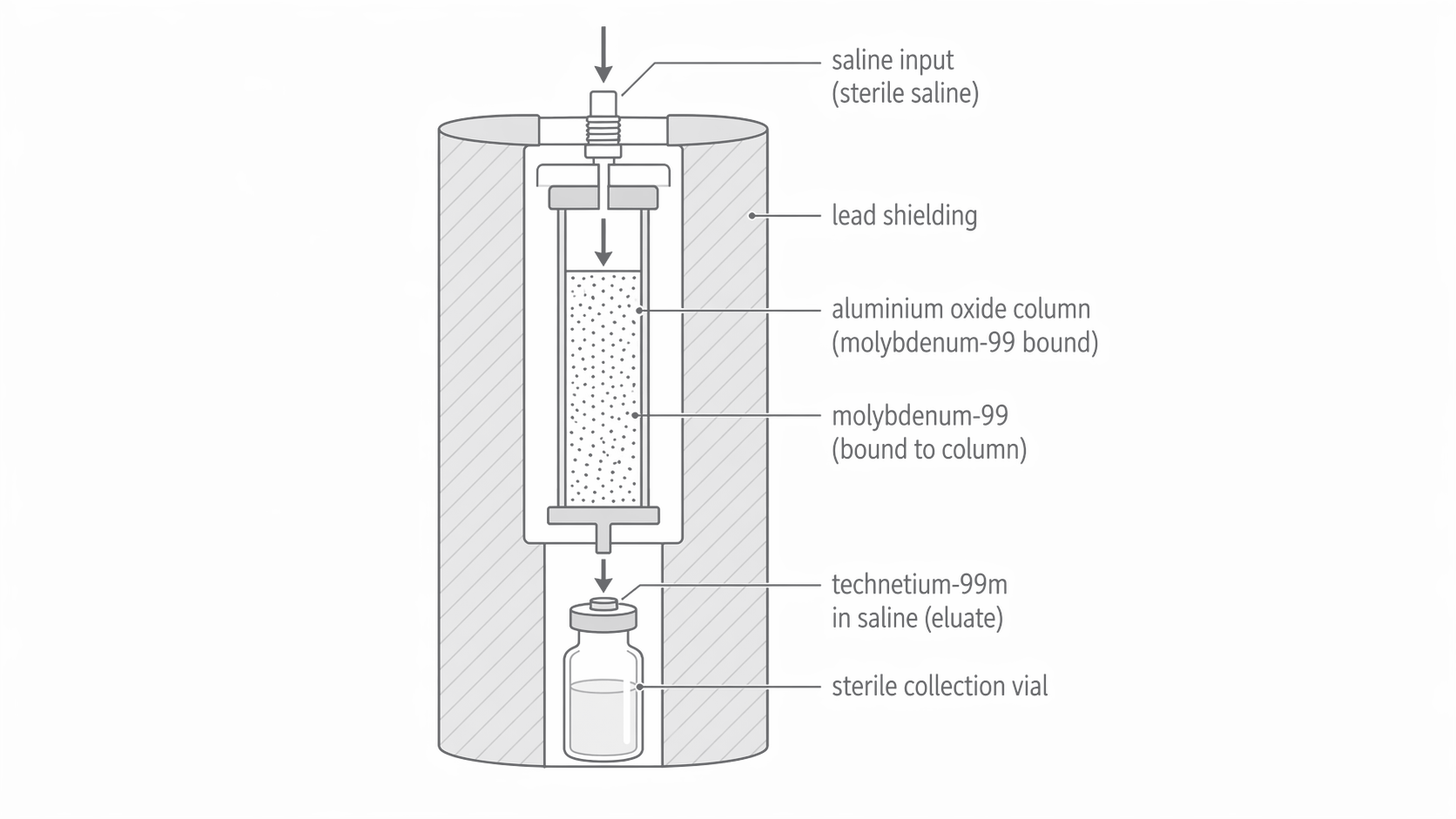
The generator does not create new radioactivity from electricity. Nuclear decay produces the daughter; chemistry separates it. Immediately after elution, daughter activity in the generator is low. It builds up again as molybdenum decays, but some newly produced technetium also decays before collection. Therefore “number extracted” is not simply all daughter nuclei ever produced. For a separated technetium sample, use technetium's decay constant in , not molybdenum's.
Continue this lesson.
You’re previewing NovaLearn without an account. Create a free account to read the rest.
Create free account