3.10.4.1 - Ultrasound Imaging
An ultrasound scanner sends short sound pulses into tissue and measures the echoes. Follow the energy conversions, calculate reflection and depth, and explain how the same signals become an A-scan trace or a B-scan image.
Generating short pulses and detecting echoes
Ultrasound is a longitudinal mechanical wave with a frequency above . In tissue, alternating compressions and rarefactions transfer energy without transporting tissue along the beam. Imaging commonly uses frequencies of a few megahertz.
A piezoelectric transducer converts between electrical signals and mechanical vibrations. An alternating potential difference across a thin piezoelectric crystal repeatedly changes its thickness. The vibrating face launches ultrasound into the adjacent material. Driving near the crystal's natural frequency produces resonance and a large vibration amplitude. Conversely, pressure variations in a returning echo deform the crystal and produce an alternating potential difference across its electrodes, which the electronics amplify.
[DIAGRAM: asset_name: B.4.1 - Ultrasound Imaging - Diagram 1; asset_slug: B.4.1 - Ultrasound Imaging - Diagram 1; caption: A piezoelectric crystal sends a pulse into tissue and converts the returning echo into an electrical signal.; description: Separate electrode leads connect to opposite crystal faces. Backing damps vibration. Pulse and echo arrows have opposite directions.]
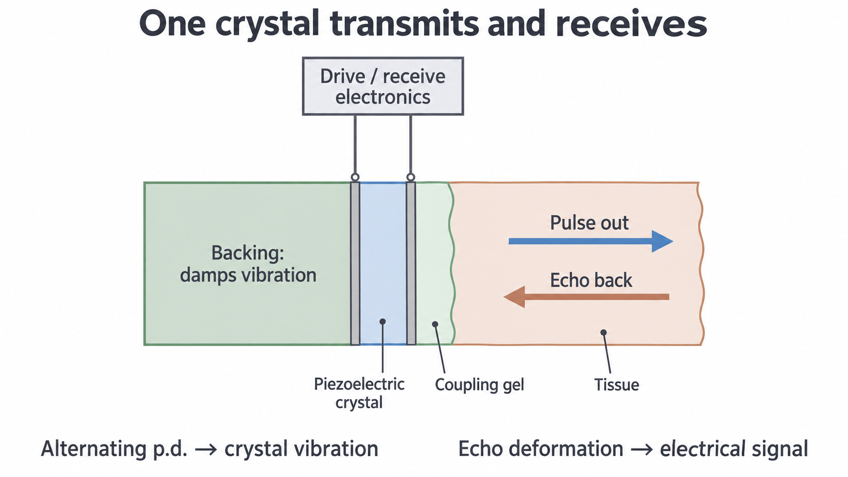
The electronics apply a short burst, then switch to receiving. Backing material damps the crystal, stopping it ringing for too long after the drive ends. This lets the same crystal detect echoes without its own vibration masking them. Short pulses also separate echoes from nearby boundaries along the beam: this is good axial resolution.
Why frequency and pulse length both matter
With sound speed and frequency , the wavelength is . A pulse containing cycles has approximate spatial length . Two reflecting boundaries need a round-trip separation at least as large as that pulse length, giving an approximate axial resolution . This estimate assumes comparable detectable echoes, and is a reasoning tool rather than a universal scanner specification.
For example, with , , and two cycles per pulse, , the pulse length is , and the estimated axial resolution is . Higher frequency shortens the wavelength, but it also usually increases attenuation in tissue, reducing useful penetration. Damping shortens the pulse without being the control that sets its frequency.
Acoustic impedance and tissue boundaries
A sound wave's behaviour at a boundary depends on both materials. The acoustic impedance of a medium is the product of its density and the speed of sound in it. It relates the pressure variation in a travelling wave to the particle velocity; it is not simply a measure of how much sound the medium absorbs.
Acoustic impedance
Density has unit , and sound speed has unit , so has unit . Acoustic impedance and attenuation are different properties: a large value of alone does not say what fraction of a wave is absorbed or reflected.
| Medium | Representative acoustic impedance / |
|---|---|
| Air | |
| Water | |
| Fat | |
| Muscle | |
| Bone | Approximately , depending on composition |
For a material with and ,
The impedance difference across a boundary, not which medium has the larger density alone, determines reflection. Similar soft-tissue impedances permit substantial transmission while still giving detectable echoes. Air and tissue have a very large mismatch.
Reflection, coupling and attenuation
At a boundary, an incident pulse is partly reflected and partly transmitted. For normal incidence, the intensity reflection coefficient is
Fraction of incident intensity reflected
and are incident and reflected intensities; and are the impedances on the two sides. The coefficient has no unit. The square is essential: the ratio inside the brackets is not the intensity fraction. Reversing the media changes its sign but not . Equal impedances give ; a very large mismatch makes approach 1.
If absorption at the interface is negligible, conservation of energy gives . This describes transmission at that boundary, before further loss while travelling through the next tissue.
Reflection is useful inside the body but undesirable at the probe
For air and tissue with and , the equation gives : about is reflected. Coupling gel removes the air gap between the probe and skin. Its impedance is close to that of tissue, so it greatly reduces reflection and allows energy into the body. It does not eliminate every internal echo; tissue-boundary echoes are what the scanner needs.
For a soft-tissue boundary with and ,
Only is reflected and approximately is transmitted at this idealised boundary. The common factor cancels. A small reflected fraction can be detected while the transmitted pulse continues to deeper boundaries.
Attenuation on the outward and return journeys
Attenuation reduces intensity as a wave travels: absorption transfers energy to the material, and scattering redirects energy away from the useful beam. A deeper echo usually travels farther both out and back, so it suffers more loss even if its reflecting boundary has the same . A swept-gain amplifier applies greater gain to later echoes to compensate. It cannot recover a signal that has fallen below the useful detection limit, nor make echo height a pure measure of impedance mismatch without accounting for other losses.
Continue this lesson.
You’re previewing NovaLearn without an account. Create a free account to read the rest.
Create free account