3.10.1.1 - Physics of Vision
The eye first forms a retinal image by refraction, then detects it with light-sensitive cells. Focusing, sensitivity, colour discrimination and resolution are different properties: a sharp optical image is useful only if the retina can detect its detail.
Refraction and the retinal image
The cornea and lens form a converging optical system. Light changes direction at boundaries between media with different refractive indices. The air–cornea boundary provides most of the eye's refraction; the lens supplies further, adjustable refraction. A ray drawing must therefore show bending at both the cornea and the lens.
[DIAGRAM: asset_name: B.1.1 - Physics of Vision - Diagram 1; asset_slug: B.1.1 - Physics of Vision - Diagram 1; caption: Structures that admit, focus and detect light in the eye; description: Structures that admit, focus and detect light in the eye]
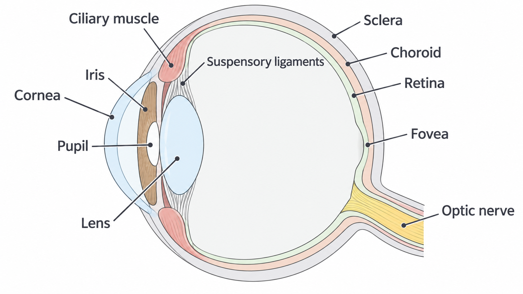
The iris changes the diameter of the pupil, the opening through which light enters. A smaller pupil admits less light and excludes some peripheral rays; it does not provide the adjustable refractive power. The retina is the detector surface. The dark choroid absorbs stray light, reducing internal reflections, while the sclera supports and protects the eye. Signals leave through the optic nerve; the exit region has no photoreceptors and is the blind spot.
Rays from one object point must meet at one retinal point. Rays from a different object point meet elsewhere, producing an image rather than one common spot for the entire scene. In ordinary viewing the retinal image is real, inverted and diminished: the rays actually converge, rays from the top of the object reach the lower retina, and the image is smaller than the object. Parallel rays represent a distant point object, not a complete extended object.
The retina is effectively fixed relative to the optical system. Unlike a camera that can move its lens or sensor, the eye focuses mainly by changing lens curvature.
Accommodation at a fixed retina
Accommodation changes the eye's optical power so that objects at different distances remain focused on the same retina. Nearer objects send more divergent rays into the eye, so a greater converging power is required.
| Viewing condition | Ciliary muscles and ligaments | Lens and optical effect |
|---|---|---|
| Distant object | Muscles relaxed, ligaments taut | Flatter lens, lower power, longer focal length |
| Near object | Muscles contract, ligament tension falls | Elastic lens becomes more curved, higher power, shorter focal length |
The muscle does not squeeze the lens directly. Reduced tension allows the lens to become rounder. Accommodation varies continuously with object distance; there is no sharp switch at a distance such as 5 m.
[DIAGRAM: asset_name: B.1.1 - Physics of Vision - Diagram 2; asset_slug: B.1.1 - Physics of Vision - Diagram 2; caption: Two-stage refraction focuses distant and near point objects on the retina; description: Two-stage refraction focuses distant and near point objects on the retina]
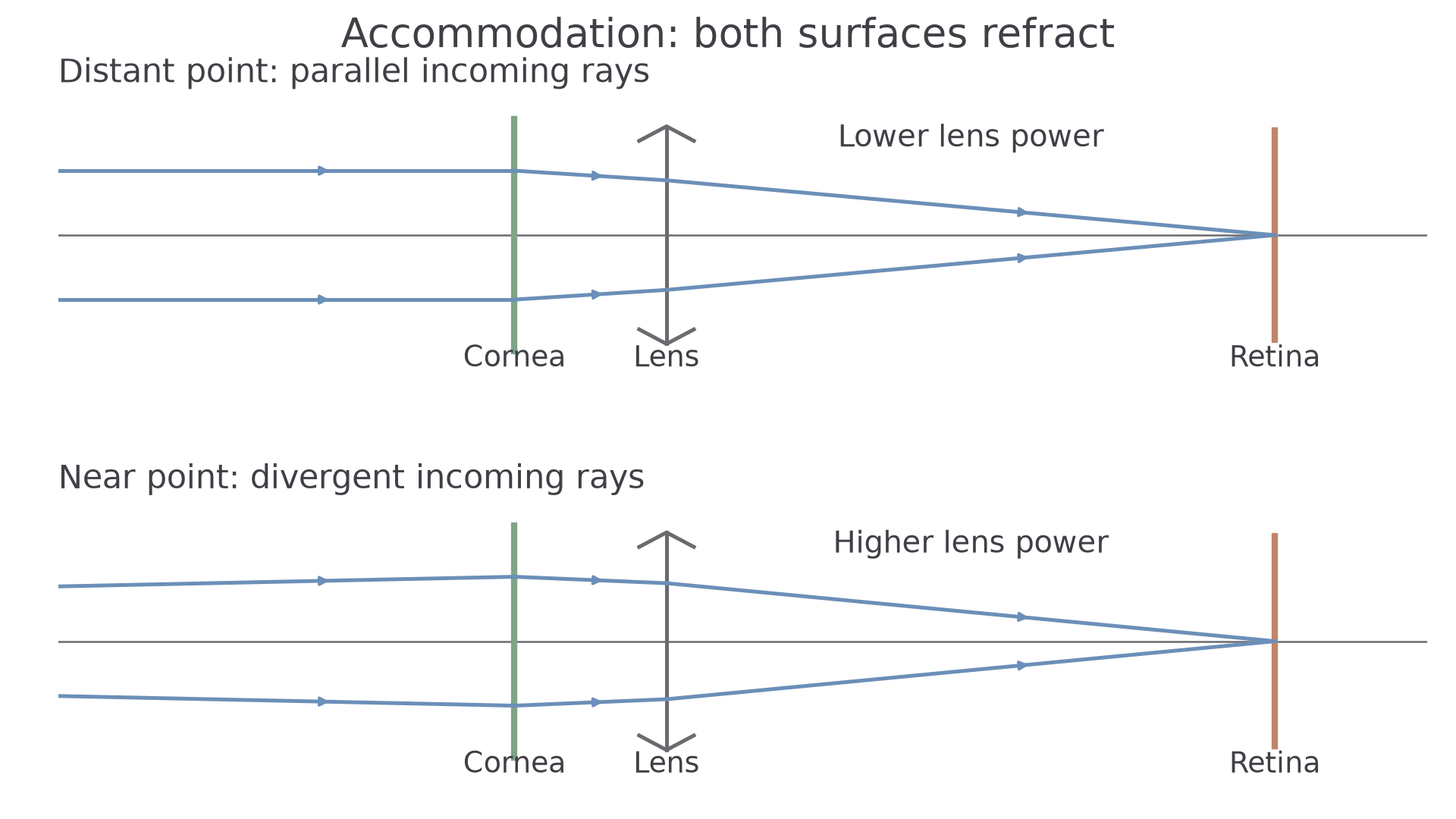
In this reduced ray model, both refracting elements bend the rays toward the axis. More lens power is needed for the divergent near-object rays. The model preserves the optical construction; its dimensions are not a scale drawing of an anatomical eye.
The far point is the furthest object position seen sharply with the eye relaxed. For an eye without a distance defect it is effectively infinity. The near point is the closest position that can be focused at maximum accommodation. The conventional normal near point used in calculations is ; actual near points depend on the person and age. Closer than the near point, even maximum power is insufficient: the rays would meet behind the retina and form a blur on it.
Rods, cones and spectral response
The retina acts as a photodetector: absorbed light changes the electrical activity of its cells, leading to nerve signals. Rods respond at low light levels. Cones need brighter conditions and provide colour vision. Both have wavelength-dependent sensitivity.
Continue this lesson.
You’re previewing NovaLearn without an account. Create a free account to read the rest.
Create free account