3.10.2.1 - Ear as a Sound Detection System
The ear transfers pressure variations in air into motion of cochlear fluid and finally into electrical signals. Follow the transfer of energy, the increase in pressure and the position-dependent response that retains information about frequency.
From air to the eardrum
The outer ear collects and guides sound. The pinna modifies the sound reaching the auditory canal and helps with directional information. Pressure variations travel down the canal and exert an alternating force on the tympanic membrane (eardrum), making it vibrate.
[DIAGRAM: asset_name: B.2.1 - Ear as a Sound Detection System - Diagram 1; asset_slug: B.2.1 - Ear as a Sound Detection System - Diagram 1; caption: Outer, middle and inner ear structures along the sound pathway; description: Outer, middle and inner ear structures along the sound pathway]
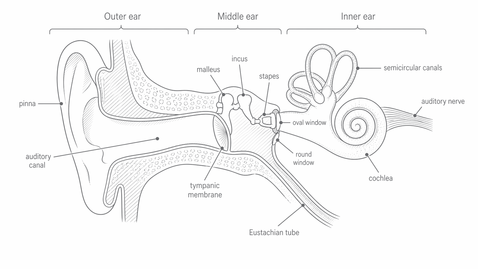
The middle ear is an air-filled cavity. The malleus, incus and stapes form the ossicle chain between the eardrum and the oval window. The inner ear contains the fluid-filled cochlea, where mechanical motion produces sensory electrical signals. The semicircular canals shown nearby serve balance, not the frequency analysis considered here.
For a steady pure tone, the forced vibrations occur at the driving frequency. A tone makes the eardrum and ossicles vibrate at ; transmitting or increasing pressure does not raise the pitch. The canal also has a resonant response that makes some frequencies more strongly represented, rather than changing them into new frequencies.
The sound path is air pressure variation → eardrum motion → ossicle motion → oval-window motion → cochlear-fluid motion → hair-cell response → nerve signal. Mechanical vibrations are not carried along the auditory nerve.
Middle-ear pressure transfer
The middle ear improves the transfer of energy from low-acoustic-impedance air to the much higher-impedance fluid of the cochlea. Direct coupling would reflect a large fraction of the incident sound. The ossicles and membrane areas act as an impedance-matching system, increasing pressure while reducing displacement; they do not create energy.
The malleus attaches to the eardrum, the incus links it to the stapes, and the stapes footplate moves the oval window. In a simplified lever model the output force is greater than the eardrum force. Use the force ratio supplied in a question; a value around 1.3–1.5 is an approximation, not a universal constant.
Because the effective eardrum area is much larger than the oval-window area, the transferred force acts over a smaller area. From ,
For an effective area ratio of 16 and a force ratio of 1.3, the pressure ratio is . This is a pressure ratio. It is not an energy gain or automatically an intensity ratio between the two different media.
For example, if the eardrum pressure amplitude is and its effective area is , its force amplitude is . A force ratio of 1.4 gives at an oval window of area : its pressure amplitude is . Squaring the millimetre-to-metre conversion matters.
Continue this lesson.
You’re previewing NovaLearn without an account. Create a free account to read the rest.
Create free account