6.1.3(e) - Electrophoresis for nucleic acids and proteins
Electrophoresis is a way of separating biological molecules by making charged molecules move through a gel in an electric field. In this lesson you are learning the principle that makes nucleic acid fragments and proteins separate, how a practical gel is run safely, and how band patterns can be used as evidence. The important habit is to say what the bands show, while avoiding claims that the evidence cannot support.
Electric Field And Gel
Electrophoresis means movement of charged particles in an electric field. In gel electrophoresis, the particles move through a gel matrix. The gel contains pores, so it acts like a molecular sieve: smaller molecules or fragments pass through the pores more easily than larger ones.
Electrophoresis
Electrophoresis is a technique that separates charged biological molecules by their movement through a medium in an electric field.
Gel matrix
A gel matrix is the porous gel through which molecules move during electrophoresis, allowing molecules with different mobility to separate.
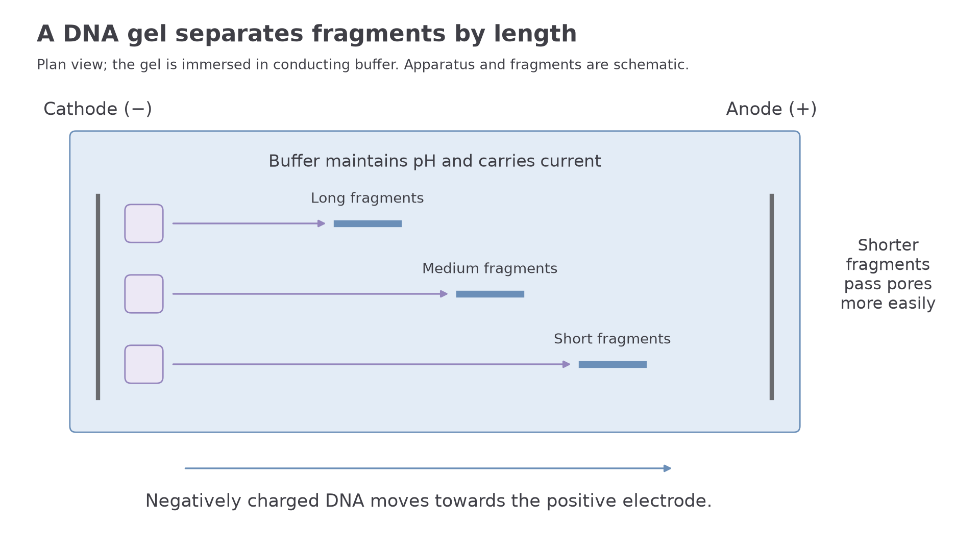
The gel sits in a buffer solution. The buffer conducts current and helps keep the pH stable. Samples are loaded into small wells at one end of the gel, then a potential difference is applied across the gel.
The electrodes matter. Negatively charged molecules move towards the positive electrode, called the anode. Positively charged molecules move towards the negative electrode, called the cathode. Nucleic acids such as DNA and RNA are negatively charged because of phosphate groups in their sugar-phosphate backbones, so they move towards the anode.
For nucleic acid fragments of the same type, shorter fragments usually travel further through the gel in a fixed time. A longer DNA fragment has more negative charges, but it also has more mass and is slowed more by the gel pores. The separation is therefore mainly by fragment length, with shorter fragments moving further from the wells.
Proteins are more complicated because their amino acid sequences give them different shapes and different net charges. A protein's movement may depend on its net charge, size and shape unless the method deliberately reduces the effect of shape and charge.
[DIAGRAM: gel-electrophoresis-migration: Lesson 152: Electrophoresis For Nucleic Acids And Proteins - diagram 01; asset_slug: 152_m06_1_3_electrophoresis_for_nucleic_acids_and_proteins__diagram_01; recommended_method: drawn_biology; description: A 16:9 drawn gel electrophoresis setup with wells near the cathode, anode at the far end, buffer, gel pores, and three negatively charged DNA fragments showing smaller fragments migrating further through the gel.]
Nucleic Acid Fragments
When electrophoresis is used for nucleic acids, the sample may contain DNA or RNA fragments of different lengths. A mixture might come from a PCR reaction, a restriction-enzyme digest, a DNA profiling investigation, or another molecular analysis. Those neighbouring techniques are not the focus here; the point is that electrophoresis separates the fragments so the pattern can be analysed.
The practical logic is simple:
| Step | Purpose |
|---|---|
| Place gel in buffer | Allows current to pass and keeps conditions stable |
| Load samples into wells | Gives each sample its own lane |
| Load a marker or ladder | Provides fragments of known length for comparison |
| Apply a potential difference | Makes charged fragments move through the gel |
| Visualise the bands safely | Shows where the separated fragments ended up |
A lane is one track through the gel from one well. A band is a visible group of many fragments that have travelled to the same position. If a sample produces two bands, it contains at least two groups of fragments with different lengths, or at least two groups that migrated differently under those conditions.
Marker or ladder
A marker, or ladder, is a sample containing fragments of known size that is run in a lane so the sizes of unknown bands can be estimated.
Interpreting DNA bands
A marker lane contains known DNA fragments of 1000 base pairs, 700 base pairs, 500 base pairs and 300 base pairs. An unknown sample has one band between the 500 base-pair and 300 base-pair marker bands, closer to the 500 base-pair band.
The unknown fragment is smaller than 500 base pairs but larger than 300 base pairs. Because it lies closer to the 500 base-pair marker band, the fragment is closer in length to 500 bp than 300 bp under an approximately logarithmic calibration, but its precise size needs measured migration distances and a calibration curve. The correct conclusion is an approximate size, not an exact sequence.
When a band in an unknown lane is at the same position as a marker band, it suggests that the unknown fragment has a similar length to that marker fragment. It does not prove that the unknown has the same base sequence. Different fragments can have the same length.
Use the direction of movement carefully. For DNA or RNA, the band furthest from the wells is usually the smallest fragment, not the largest.
Protein Electrophoresis
Electrophoresis can also separate proteins. This is useful because cells often contain mixtures of many proteins, and a researcher may want to compare protein samples, estimate the relative molecular mass of a protein, or check whether a purification step has removed other proteins.
The same general idea applies: molecules move in an electric field and the gel slows them. The difference is that proteins do not all have the same charge pattern. Their side chains can be positive, negative, polar or non-polar, and the folded shape of a protein can affect how easily it passes through the gel.
In native protein electrophoresis, proteins keep more of their natural structure. Their mobility can depend on net charge, size and shape. That can be useful for some comparisons, but it makes simple size interpretation harder.
A common protein method is SDS-PAGE. SDS is a detergent that denatures many proteins and gives them a more uniform negative charge relative to their mass. In that method, the proteins move towards the positive electrode and separate mainly by molecular mass. Smaller polypeptides usually move further through the gel than larger polypeptides.
Continue this lesson.
You’re previewing NovaLearn without an account. Create a free account to read the rest.
Create free account