3.1.2(c)-(d) - Blood vessels, tissue fluid and lymph
Blood is useful only if it can be moved close enough to cells for exchange to happen. In this lesson you will learn how the structure of arteries, arterioles, capillaries, venules and veins matches their function, then use pressure differences to explain how tissue fluid and lymph form. The key exam skill is to describe movement of fluid using hydrostatic pressure and oncotic pressure, not vague phrases such as "blood leaks into tissues".
The Route Through Blood Vessels
In a mammalian circulation, blood does not pass straight from a large artery into a large vein. It moves through a branching sequence:
arteries -> arterioles -> capillaries -> venules -> veins
Each vessel type solves a different problem. Arteries carry blood away from the heart under high pressure. Arterioles adjust the supply of blood to capillary beds. Capillaries are the exchange surface between blood and body cells. Venules collect blood from capillaries. Veins return blood to the heart at low pressure.
Do not define arteries as "oxygenated" and veins as "deoxygenated". That shortcut fails in the pulmonary circulation. The definitions are based on direction of flow: arteries carry blood away from the heart; veins carry blood towards the heart.
| Vessel type | Main function | Structure that matches the function |
|---|---|---|
| Artery | Carries blood away from the heart under high pressure | Thick wall, much elastic tissue, much smooth muscle, collagen for strength, relatively narrow lumen |
| Arteriole | Controls blood flow into capillary beds | Smooth muscle can constrict or dilate the lumen, changing resistance and blood supply |
| Capillary | Allows exchange between blood and tissue fluid | Very thin wall made from endothelium, very narrow lumen, large total surface area, close to cells |
| Venule | Collects blood from capillaries | Thin wall and wider lumen than capillaries; pressure is already low |
| Vein | Returns blood to the heart at low pressure | Thin wall, large lumen, less muscle and elastic tissue than arteries, valves to prevent backflow |
[DIAGRAM: vessel_wall_comparison: Lesson 060: Blood vessels, tissue fluid and lymph - diagram 01; asset_slug: 060_m03_1_2_blood_vessels_tissue_fluid_and_lymph__diagram_01; recommended_method: drawn_biology; description: labelled comparison of artery, arteriole, capillary, venule and vein cross-sections showing lumen size, wall thickness, endothelium, smooth muscle, elastic tissue, collagen and a venous valve.]
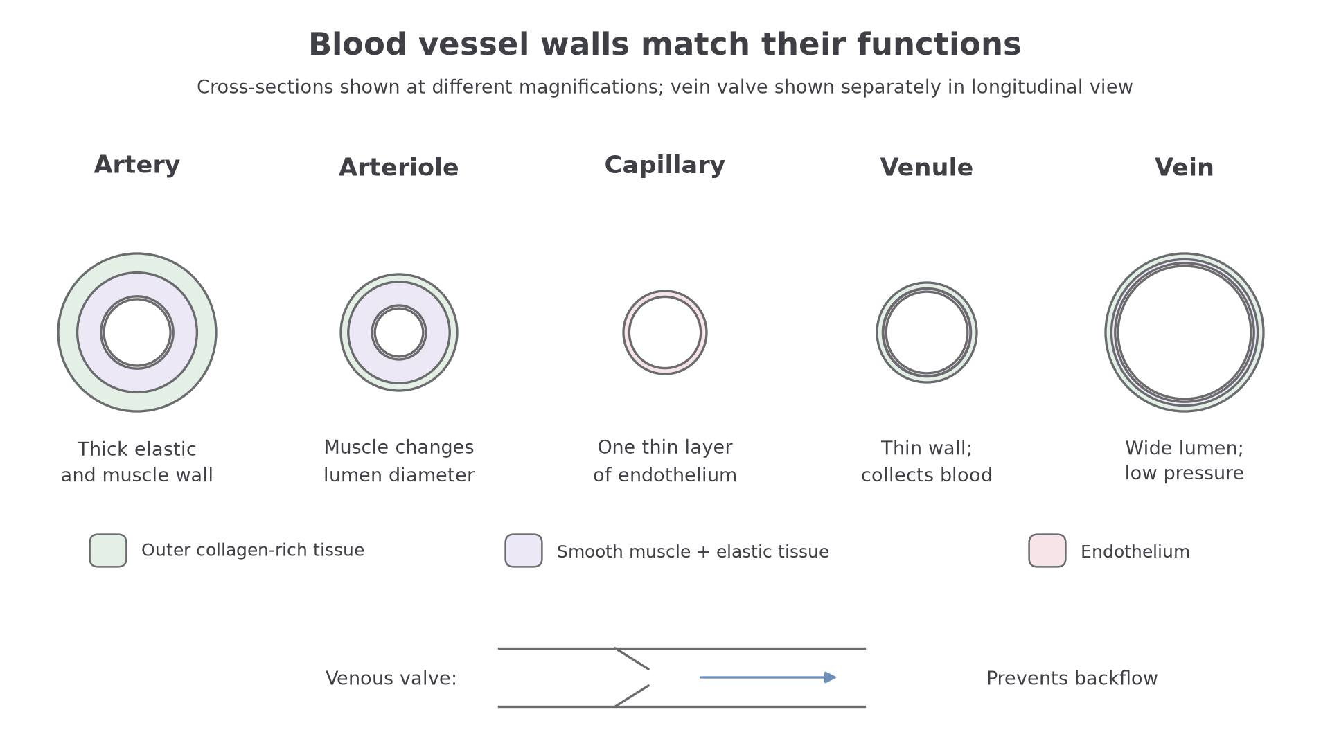
The comparison matters because pressure falls as blood travels further from the heart and through narrower resistance vessels. High pressure needs strong, elastic walls. Exchange needs a short diffusion distance. Low-pressure return needs a large lumen and help from valves.
Wall Tissues And Practical Recognition
OCR expects you to know not just that vessel walls are "thick" or "thin", but how different tissues are distributed within the wall.
The innermost lining is endothelium. It is a thin, smooth layer of flattened cells that reduces friction and forms the barrier between blood and the vessel wall. Capillaries are mainly endothelium, so substances have only a short distance to move between blood plasma and tissue fluid.
Around the endothelium, arteries and arterioles have much more smooth muscle and elastic tissue than veins. Smooth muscle can contract, narrowing the lumen. This is especially important in arterioles because changing arteriole diameter changes resistance and therefore controls how much blood enters a capillary bed. Elastic tissue stretches when pressure rises and recoils when pressure falls, helping to maintain blood flow between heartbeats.
The outer part of larger vessel walls contains collagen-rich connective tissue. Collagen gives tensile strength, so vessels are less likely to burst or over-stretch. Veins have some smooth muscle, elastic tissue and collagen, but much less than arteries. Their lower pressure means they do not need such thick elastic walls. Many veins also contain valves made from folds of the endothelium, reinforced by connective tissue.
In a microscope image or dissection context, identify an artery by its thick wall and relatively small, rounded lumen. Identify a vein by its thinner wall, wider often irregular lumen, and possible valves. A capillary may not be visible as a neat tube in a low-power section because it is only about one endothelial cell thick.
PAG2 links this content to dissection and observation. You are not expected in this lesson to learn a full heart dissection method, but you should be able to connect safe practical observation to the biology: use sharp instruments carefully, keep animal tissue hygienic, observe sections systematically, and use labelled scientific drawings to show wall thickness, lumen shape and visible tissue layers.
The vessel wall is not just a pipe. Its endothelium, smooth muscle, elastic tissue, collagen and valves are distributed differently because each vessel type experiences a different pressure and has a different job.
Formation Of Tissue Fluid
Body cells are not usually in direct contact with blood. The immediate fluid around most body cells is tissue fluid. It forms from blood plasma at capillary beds and acts as the exchange medium between capillaries and cells.
At the arteriole end of a capillary bed, blood hydrostatic pressure is relatively high. Hydrostatic pressure is the pressure exerted by a fluid against a surface. In this context, it pushes plasma out through the thin capillary wall. Water and small dissolved substances such as oxygen, glucose, amino acids, ions and hormones can leave the blood and enter the spaces around cells.
Red blood cells, platelets and most plasma proteins remain in the capillary. They are too large to pass through the capillary wall easily. This is important because retained plasma proteins create oncotic pressure.
Oncotic Pressure
Oncotic pressure is the inward-pulling pressure caused mainly by plasma proteins that remain in the blood and attract water back towards the capillary.
In the simplified capillary-pressure model used for these OCR-style questions, hydrostatic pressure falls along the capillary while retained proteins sustain an inward oncotic effect. If that inward effect exceeds the outward pressure near the venule end, net movement can be back into the capillary. This is a conditional model, not a fixed proportion of fluid returning in every tissue: actual exchange also depends on permeability and the full pressure balance, and lymphatic return is important. Dissolved carbon dioxide and wastes can move from cells towards blood.
Continue this lesson.
You’re previewing NovaLearn without an account. Create a free account to read the rest.
Create free account