2.1.2(n)-(o) - Globular and fibrous proteins
This lesson is about how protein shape fits biological function. You already met amino acids, peptide bonds and levels of protein structure; here the focus narrows to two broad protein types that OCR names directly: globular proteins and fibrous proteins. You will use haemoglobin, a named enzyme and insulin for globular proteins, then collagen, keratin and elastin for fibrous proteins.
Shape and function
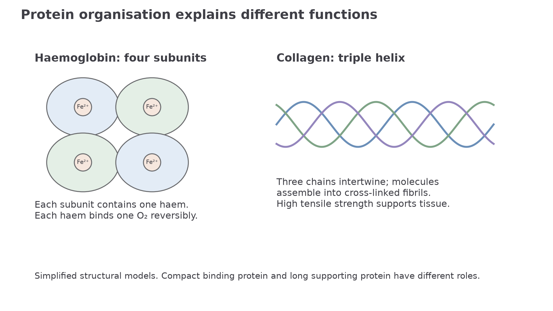
[DIAGRAM: globular_fibrous_models: Lesson 025: Haemoglobin and collagen; asset_slug: 025_m02_1_2_globular_and_fibrous_proteins__diagram_01; recommended_method: drawn_biology; description: Four haem-containing globin subunits and a collagen triple helix relate structure to transport and support.]
Proteins are not useful because they are "made of amino acids" in a general way. They are useful because their amino acid sequence folds into a three-dimensional shape that gives a particular property or job.
Globular proteins fold into compact, roughly rounded molecules. They often have hydrophilic regions on the outside, so they can interact with water and dissolve or move in watery environments such as cytoplasm or blood plasma. Their specific tertiary or quaternary shape lets them bind other molecules, act as enzymes, transport substances, or work as signalling molecules.
Fibrous proteins are longer, less compact proteins used mainly for structural roles. For this specification point, you only need their properties and functions, not detailed molecular structures. The key idea is that fibrous proteins tend to be tough, insoluble and mechanically useful in tissues.
Ask whether the protein's role depends mainly on specific molecular binding or on mechanical properties such as strength and elasticity.
Globular proteins are usually compact and specific; fibrous proteins are usually structural and mechanically useful.
Haemoglobin as a conjugated protein
Each of haemoglobin’s four subunits contains a haem prosthetic group with an iron(II) ion, Fe²⁺. Each haem can bind one oxygen molecule reversibly, so one haemoglobin molecule can carry four O₂ molecules. Reversible binding allows loading where oxygen is abundant and unloading in respiring tissue; permanent binding would prevent transport. The four haem groups are non-protein components, whereas the four globin chains provide quaternary structure.
Haemoglobin is OCR's required example of a conjugated protein. A conjugated protein is a protein with a non-protein part attached or associated with it. That non-protein part is called a prosthetic group.
Prosthetic group
A non-protein component that is permanently associated with a protein and is needed for the protein's function.
Haemoglobin is a globular protein with quaternary structure. It contains four polypeptide subunits, and each subunit is associated with a haem prosthetic group. The haem group contains an iron ion that is involved in binding oxygen. For this lesson, the key point is not the full oxygen dissociation curve. The key point is that haemoglobin's protein part and non-protein haem groups work together, so haemoglobin is a conjugated globular protein.
Interpreting the phrase conjugated protein
If a question says haemoglobin is a conjugated protein, the answer should link two ideas:
- haemoglobin is a protein made of polypeptide chains
- it also contains a non-protein prosthetic group, haem, needed for its function
A strong answer would be: "Haemoglobin is conjugated because it is a globular protein with haem prosthetic groups associated with its polypeptide subunits."
Do not overclaim from the word "globular". Globular does not mean the molecule is a perfect sphere, and it does not mean its shape never changes. It means the overall protein is compact rather than long and fibrous.
Enzymes and insulin
Insulin has two polypeptide chains linked by disulfide bridges. Its folded surface binds a specific receptor on a target-cell membrane, initiating a response that helps lower blood glucose. It does not act as an enzyme that breaks down glucose. A soluble hormone can travel in blood, whereas its specific shape determines which cells respond.
A named enzyme is another required globular-protein example. This lesson will use amylase. Amylase is an enzyme that catalyses the breakdown of starch into smaller sugars. Its folded globular shape creates an active site: a region with a specific shape and chemical environment that binds the substrate.
Active site
The region of an enzyme where the substrate binds and the reaction is catalysed.
The structure-function link for a named enzyme is therefore:
| Feature of a globular enzyme | Why it matters |
|---|---|
| Specific folded tertiary structure | Creates the active site shape and chemical environment |
| Hydrophilic regions on the outside | Helps many enzymes work in watery cell or body-fluid environments |
| Bonds and interactions maintaining shape | Keep the active site in a functional arrangement |
Insulin is also a globular protein. Its function is signalling: it is a hormone that can bind to specific receptors on target cells. In this specification point, you only need insulin as an example of a globular protein whose shape is important for function. Detailed control of blood glucose, secretion from pancreatic beta cells and diabetes treatment belong to later lessons.
Choosing the right structure-function link
Suppose a question asks why a change in the tertiary structure of amylase could stop it working.
A weak answer is: "The enzyme changes shape."
A stronger answer is: "The active site changes shape, so starch is less able to bind correctly and the enzyme-substrate complex forms less often."
For insulin, the equivalent reasoning is about receptor binding rather than catalysis. If the shape of insulin is altered enough, it may bind less effectively to its receptor, so the signal is not received normally by the target cell.
Fibrous examples
Fibrous proteins are mainly useful because of their physical properties. OCR names three examples: collagen, keratin and elastin. You must know what properties they have and what functions those properties support. You do not need details of their molecular structures.
| Fibrous protein | Useful property | Function examples |
|---|---|---|
| Collagen | Strong and resistant to stretching | Provides tensile strength in tendons, skin, blood vessel walls, cartilage and bone matrix |
| Keratin | Tough, insoluble and protective | Helps form hair, nails and the outer layer of skin |
| Elastin | Elastic: can stretch and recoil | Allows tissues such as skin, lungs and blood vessel walls to return towards their original shape |
Collagen is the example to reach for when a question is about tensile strength. Tensile strength means resisting pulling forces. That is why collagen-rich tissues can hold structures together and withstand tension.
Keratin is the example to reach for when a question is about toughness or protection. Hair and nails are not trying to transport substances or catalyse reactions; they are protective structures.
Elastin is the example to reach for when a question is about stretch and recoil. A blood vessel wall, for example, needs to stretch as pressure changes and then recoil rather than staying stretched.
If a question asks about fibrous proteins in this specification point, do not spend marks on collagen triple helices, keratin coiled coils or elastin cross-links. The rewarded focus is properties and functions.
A safe fibrous-protein answer should name the protein, state the property, and link that property to the tissue function.
Compare and apply
A compare answer must refer to both protein types throughout. Do not write one paragraph about globular proteins and then a separate list about fibrous proteins if the question asks for direct differences.
| Comparison point | Globular proteins | Fibrous proteins |
|---|---|---|
| Overall shape | Compact, folded, roughly rounded | Long or fibre-like |
| Typical solubility | Often soluble in water | Usually insoluble |
| Main role | Specific binding, catalysis, transport or signalling | Structural support, strength, protection or elasticity |
| Examples in this lesson | Haemoglobin, amylase, insulin | Collagen, keratin, elastin |
Building a compare answer
Question: Compare haemoglobin and collagen as examples of globular and fibrous proteins.
A good answer might say:
"Haemoglobin is a compact globular protein, whereas collagen is a fibrous protein. Haemoglobin is soluble enough to function in red blood cells and contains haem prosthetic groups for oxygen binding, whereas collagen provides tensile strength in tissues. Haemoglobin's function depends on specific binding, while collagen's function depends mainly on mechanical support."
Notice that each sentence compares both examples or links a feature directly to a function.
One common misconception is that "fibrous" means "less biological" or "less specific". Fibrous proteins are just as precisely made by cells. The difference is the kind of useful outcome: mechanical properties rather than a compact binding site or signalling shape.
Models and boundaries
To investigate a protein model, select one chain and trace its backbone from one end to the other; then colour other chains differently. Identify helices or sheets, display cysteine side chains to locate disulfide links, and locate any non-protein group separately. Compare haemoglobin’s four compact subunits with collagen’s three intertwined chains. A ribbon model shows overall folding clearly but omits many atoms; a space-filling model shows occupied volume but hides internal bonds. Record which inference each representation supports rather than treating either model as a photograph. This is preparation for PAG10; manipulating and interpreting the model is the practical activity.
PAG10 links this specification point to computer modelling or software use. In this lesson, a useful modelling task would be to use a reliable protein-structure database or molecular viewer to inspect a protein such as haemoglobin, insulin or amylase.
A good model-based investigation has a clear question, such as:
| Modelling question | What the model can help show | What the model cannot prove by itself |
|---|---|---|
| Is this protein compact or extended? | Overall shape and chain arrangement | Whether the protein works in a living cell under all conditions |
| Does haemoglobin include a non-protein group? | Haem groups can be displayed separately from polypeptide chains | Full oxygen transport physiology |
| Where might an enzyme bind substrate? | A cleft or active-site region may be visible | The full enzyme mechanism and rate |
The practical skill is not just clicking around a model. You should use a named reliable source, record which protein or structure entry you used, describe the feature you observed, and avoid conclusions that go beyond the evidence. For example, seeing a haem group in a haemoglobin model supports the statement that haemoglobin is conjugated, but it does not replace experimental evidence about oxygen dissociation.
The boundary of this lesson matters. Haemoglobin transport curves, detailed enzyme action, and blood glucose regulation are all important biology, but they are not the rewarded centre of this specification point. Here, the centre is protein type, property and function.