3.1.1(c)-(d) - Mammalian gaseous exchange system and ventilation
Mammals need a system that gets fresh air to a thin exchange surface and then keeps replacing that air. In this lesson, you will learn the route air takes through the mammalian gaseous exchange system, how the main tissues in the airways support their functions, and how muscle action changes thoracic volume and pressure during ventilation. The key is to connect structure to function instead of memorising disconnected labels.
Route to alveoli
Air entering a mammal does not exchange gases equally at every point in the system. Most of the branching tubes are conducting airways: they carry air, keep it moving, and help protect the delicate exchange surface. The main exchange surface is the alveoli, which are tiny air sacs at the end of the airway tree.
The route for this lesson is:
trachea → bronchi → bronchioles → alveoli
The trachea is the wide airway leading from the throat region towards the lungs. It divides into two bronchi, one entering each lung. Each bronchus branches repeatedly into smaller bronchioles. The smallest bronchioles lead towards clusters of alveoli, where oxygen diffuses into the blood and carbon dioxide diffuses from the blood into the air spaces.
Ventilation
The movement of air into and out of the lungs.
Ventilation matters because diffusion depends on concentration gradients. If air in the alveoli were not replaced, oxygen concentration in the alveolar air would fall and carbon dioxide concentration would rise. Moving air in and out helps maintain a high oxygen concentration and a low carbon dioxide concentration in the alveoli compared with the blood arriving at the exchange surface.
Do not blur ventilation with respiration. Ventilation is a physical movement of air. Respiration is a cellular metabolic process that releases energy in cells. Ventilation supports aerobic respiration by helping oxygen enter the body and carbon dioxide leave it, but the two words do not mean the same thing.
The airway tree is arranged so air is conducted through supported, protected tubes to thin, elastic alveoli where diffusion can occur.
Component distribution
The mammalian gaseous exchange system works because different tissues appear in different parts of the airway tree. A strong answer should say both where a component is found and what it does there.
[DIAGRAM: airway_component_distribution: Lesson 054: Mammalian gaseous exchange system and ventilation - diagram 01; asset_slug: 054_m03_1_1_mammalian_gaseous_exchange_system_and_ventilation__diagram_01; recommended_method: drawn_biology; description: Distribution of airway tissues for cartilage, ciliated epithelium, goblet cells, smooth muscle and elastic fibres across trachea, bronchi, bronchioles and alveoli.]
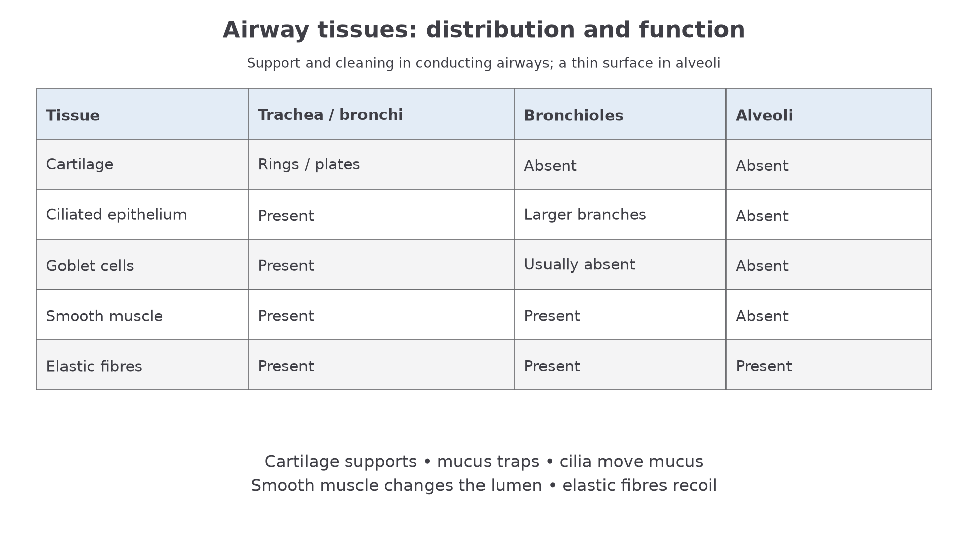
Cartilage is strong but flexible connective tissue. In the trachea it forms C-shaped rings, and in bronchi it appears as smaller plates. Its function is to support the airway and prevent collapse when pressure inside the thorax changes during breathing. Alveoli do not have cartilage because the exchange surface must stay thin and flexible.
Goblet cells secrete mucus. Mucus traps dust, bacteria and other particles before they reach the alveoli. Ciliated epithelium has cells with tiny moving cilia. The cilia move the mucus towards the throat, where it can be swallowed or removed. The goblet cells make the mucus; the ciliated cells move it. Mixing those functions is a common error.
Smooth muscle is found in the airway walls, especially around bronchi and bronchioles. When it contracts, it narrows the airway lumen. When it relaxes, the lumen widens. This helps control airflow to different parts of the lung. If smooth muscle contracts too strongly, airflow is restricted.
Elastic fibres stretch when air enters and recoil when air leaves. Elastic tissue is especially important in alveoli because recoil helps push air out during normal expiration. Elastic fibres also help the tissues return to their original shape after being stretched.
The main distribution pattern is: trachea and bronchi have cartilage, ciliated epithelium, goblet cells, smooth muscle and elastic fibres. Bronchioles lack cartilage and contain smooth muscle and elastic fibres. Ciliated epithelium occurs in larger bronchioles; goblet cells are usually absent and the smallest airways do not have the mucus-producing lining of the trachea. The distribution table is a simplified course pattern rather than a rule for every transitional airway section. Alveoli have elastic fibres and a very thin wall for exchange, but not cartilage, ciliated epithelium or goblet cells.
Alveoli and exchange surface
Alveoli are the end point of the pathway, but they are not just "small tubes". They are air sacs adapted for diffusion. Their walls are very thin, and each alveolus lies close to blood capillaries. That creates a short diffusion pathway between the air and the blood.
Elastic fibres give alveoli two useful properties. First, alveoli can stretch as air enters during inspiration. Second, they recoil during expiration, helping reduce lung volume and push air out. This recoil is one reason normal expiration can be passive.
Alveoli should not contain the airway-cleaning features of the larger tubes. Cilia and mucus would increase the diffusion pathway and interfere with gas exchange. Cartilage would make the walls too thick and rigid. The protection job is mainly done earlier in the conducting airways, so the alveoli can remain thin and specialised for diffusion.
For structure-function questions, link the structure to the process. "Elastic fibres are present" is weaker than "elastic fibres stretch during inspiration and recoil during expiration, helping air leave the alveoli."
The exchange surface still depends on ventilation. Fresh air entering the alveoli keeps oxygen concentration relatively high. Air leaving removes carbon dioxide-rich air. This maintains concentration gradients for oxygen and carbon dioxide across the alveolar wall.
Continue this lesson.
You’re previewing NovaLearn without an account. Create a free account to read the rest.
Create free account