5.1.3(d) - Synapses and neurotransmission
Neurones carry action potentials along their membranes, but one neurone usually has to pass information to another cell across a tiny gap. In this lesson you will learn how a cholinergic synapse uses acetylcholine to transmit a signal, and how synapses help decide whether a postsynaptic neurone fires. The key idea is not just "a message crosses a gap": synapses convert, filter and integrate nervous information.
Synapse As A Junction
A synapse is a junction between two cells where information is passed from a presynaptic cell to a postsynaptic cell. In this lesson the cells are neurones, although synapses can also connect neurones to effector cells such as muscle cells.
Presynaptic And Postsynaptic
The presynaptic neurone is the neurone before the synapse that releases neurotransmitter. The postsynaptic neurone is the neurone after the synapse that has receptors for the neurotransmitter.
The narrow space between the two cell membranes is the synaptic cleft. Be precise with that term. A synapse is the whole junction, including the presynaptic knob, the cleft and the postsynaptic membrane. The synaptic cleft is only the gap.
Synapses matter because an electrical signal cannot simply continue as the same action potential across the gap. Instead, the presynaptic neurone converts electrical activity into a chemical signal called a neurotransmitter. The neurotransmitter diffuses across the synaptic cleft and affects the postsynaptic membrane.
Neurotransmitter is stored in vesicles on the presynaptic side before release; the postsynaptic membrane carries the receiving receptors. This asymmetry helps identify the sides in a micrograph.
This conversion gives synapses several roles:
| Role of synapses | What that means |
|---|---|
| Direction | Transmission usually goes from the presynaptic neurone to the postsynaptic neurone because release machinery is on one side and receptors are on the other. |
| Integration | A postsynaptic neurone can receive signals from many synapses and combine their effects. |
| Filtering | Weak or scattered inputs may not trigger a new action potential; stronger combined inputs may reach threshold. |
| Control | Some synapses make firing more likely, while others make firing less likely. |
Chemical transmission introduces a synaptic delay: channels must open, vesicles fuse, transmitter diffuse and receptors bind. These stages take time compared with electrical spread over the same short distance along an axon. Adding synapses to a pathway therefore adds delays as well as opportunities to integrate information.
Cholinergic Synapse Structure
A cholinergic synapse is a synapse that uses acetylcholine, often shortened to ACh, as its neurotransmitter. OCR names this synapse type because it gives a clear example of neurotransmitter action.
[DIAGRAM: cholinergic_synapse_structure: Lesson 105: Synapses and neurotransmission - diagram 01; asset_slug: 105_m05_1_3_synapses_and_neurotransmission__diagram_01; recommended_method: drawn_biology; description: Labelled drawn biology schematic of a cholinergic synapse showing presynaptic knob, synaptic vesicles containing acetylcholine, voltage-gated calcium ion channels, synaptic cleft, acetylcholinesterase, postsynaptic receptors and sodium ion entry.]
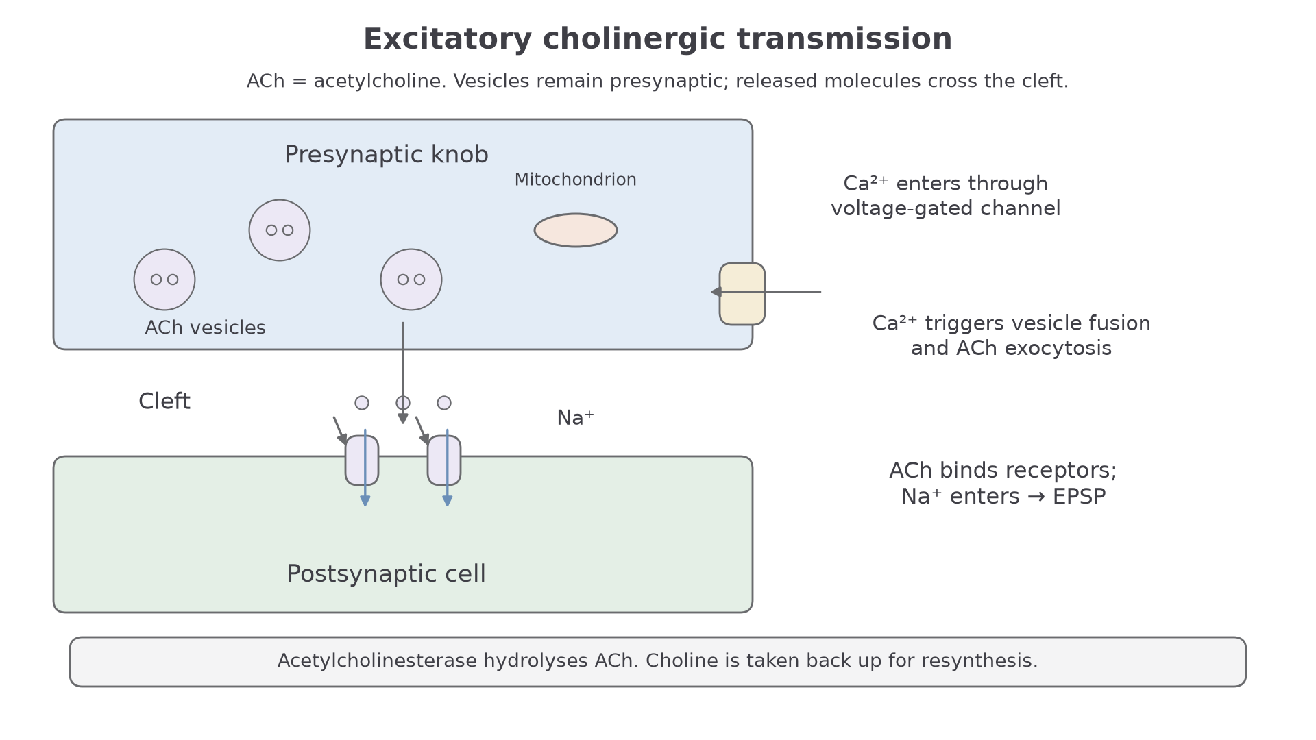
The presynaptic knob is the swollen end of the presynaptic neurone. It contains synaptic vesicles filled with acetylcholine. It also contains mitochondria, which supply ATP for processes such as vesicle movement and neurotransmitter recycling.
The presynaptic membrane contains voltage-gated calcium ion channels. These open when an action potential arrives. The postsynaptic membrane contains receptor proteins that are complementary to acetylcholine. In a simple excitatory cholinergic synapse, these receptors are associated with sodium ion channels, so acetylcholine binding can increase sodium ion permeability.
| Structure | Role in neurotransmission |
|---|---|
| Presynaptic knob | Contains vesicles and releases neurotransmitter. |
| Synaptic vesicles | Store acetylcholine before release. |
| Voltage-gated calcium ion channels | Open when the presynaptic membrane is depolarised. |
| Synaptic cleft | Gap across which acetylcholine diffuses. |
| Postsynaptic receptors | Bind acetylcholine and cause ion channels to open. |
| Acetylcholinesterase | Breaks down acetylcholine in the cleft so the signal is not continuous. |
The arrangement also explains direction. Vesicles containing acetylcholine are on the presynaptic side, while the matching receptors are on the postsynaptic side. A common error is to say that vesicles travel across the cleft. They do not. Vesicles fuse with the presynaptic membrane and release acetylcholine; acetylcholine molecules then diffuse across the cleft.
Acetylcholine Transmission
At an excitatory cholinergic synapse, each event causes the next: electrical activity controls transmitter release, and transmitter binding changes the postsynaptic membrane.
- An action potential arrives at the presynaptic knob.
- Depolarisation of the presynaptic membrane opens voltage-gated calcium ion channels.
- Calcium ions diffuse into the presynaptic knob.
- Calcium ions cause synaptic vesicles to move to and fuse with the presynaptic membrane.
- Acetylcholine is released into the synaptic cleft by exocytosis.
- Acetylcholine diffuses across the synaptic cleft.
- Acetylcholine binds to complementary receptors on the postsynaptic membrane.
- Sodium ion channels open, sodium ions diffuse into the postsynaptic neurone, and the postsynaptic membrane depolarises.
- If depolarisation reaches threshold, a new action potential is generated in the postsynaptic neurone.
Explaining a blocked calcium channel
Suppose a toxin blocks voltage-gated calcium ion channels in the presynaptic membrane.
If calcium ions cannot enter the presynaptic knob, vesicles are much less likely to fuse with the presynaptic membrane. Less acetylcholine is released into the synaptic cleft, so fewer receptors on the postsynaptic membrane are activated. The postsynaptic membrane is less likely to depolarise to threshold, so synaptic transmission is reduced or stops.
Continue this lesson.
You’re previewing NovaLearn without an account. Create a free account to read the rest.
Create free account