5.1.2(c)(i)-(iii) - Kidney structure, nephrons and practicals
The kidney is a homeostatic organ because it removes nitrogenous waste from the blood while conserving useful substances and helping the body produce urine with the right composition. In this lesson, you will connect the gross structure seen in a kidney dissection to the microscopic structure of nephrons. You will then use that structure to explain ultrafiltration, selective reabsorption, urine production and the practical skills needed to examine and draw kidney tissue.
Gross Kidney Structure
A mammalian kidney receives blood through the renal artery and returns blood through the renal vein. It is not just a waste-removal bag: it is a highly organised organ with regions that support filtration, reabsorption and urine drainage.
[DIAGRAM: gross_kidney_dissection_map: Lesson 098: Kidney structure, nephron action and kidney practicals - diagram 01; asset_slug: 098_m05_1_2_kidney_structure_nephron_action_and_kidney_practicals__diagram_01; recommended_method: drawn_biology; description: A clean line diagram comparing the external kidney with a longitudinal cut face, labelled with capsule, cortex, medulla pyramids, renal pelvis, ureter, renal artery, renal vein and hilum for dissection/drawing.]
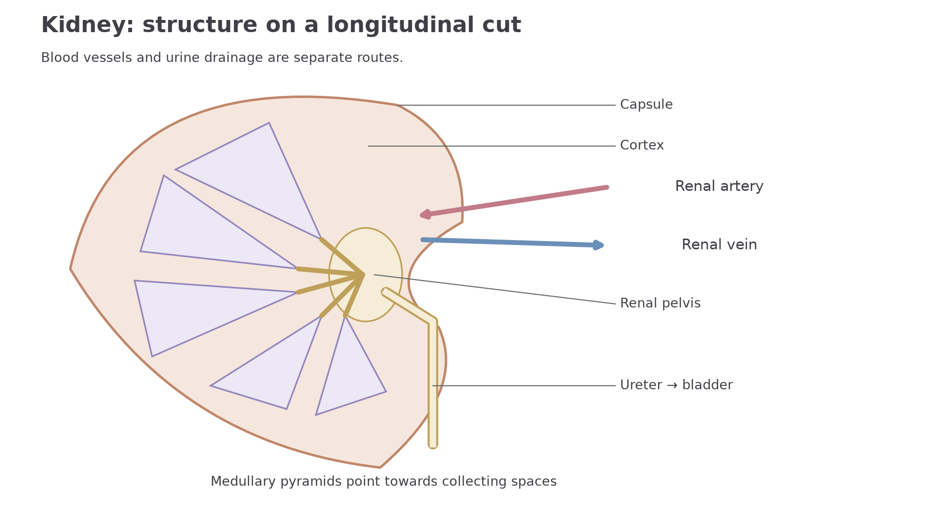
The tough outer capsule protects the kidney. Just inside it is the cortex, where many renal corpuscles and convoluted tubules are found. Deeper inside is the medulla, which contains medullary pyramids, loops of Henle and collecting ducts. The renal pelvis is the central collecting space that receives urine and leads into the ureter.
The hilum is the indentation where major vessels and the ureter enter or leave. In a dissection or drawing, this is a useful orientation point because the renal artery, renal vein and ureter are all associated with it.
Excretion
Excretion is the removal of metabolic waste products from the body.
In this lesson, the main nitrogenous waste is urea. Urea is made in the liver and transported in the blood to the kidneys. The kidney must remove enough urea while retaining useful molecules such as glucose, amino acids, many ions and most water.
This lesson is about kidney structure and nephron action. The detailed hormonal control of water potential by ADH, kidney failure treatments, and diagnostic urine tests are neighbouring lessons.
Nephron Layout And Blood Supply
The nephron is the functional unit of the kidney. Each nephron is a microscopic tubule associated with blood vessels, and each part has a different role in turning blood plasma into urine.
Nephron
A nephron is a kidney tubule and its associated blood vessels that filters blood and modifies the filtrate to form urine.
The nephron begins with the renal corpuscle. This is made from the glomerulus, a knot of capillaries, surrounded by Bowman's capsule. Blood enters the glomerulus through the afferent arteriole and leaves through the efferent arteriole. The efferent arteriole is narrower, helping to maintain the high pressure needed for filtration.
[DIAGRAM: nephron_action: Lesson 098: Nephron layout - diagram 02; asset_slug: 098_m05_1_2_kidney_structure_nephron_action_and_kidney_practicals__diagram_02; recommended_method: drawn_biology; description: Continuous filtrate route through cortex and medulla, separate from afferent, efferent and peritubular blood vessels.]
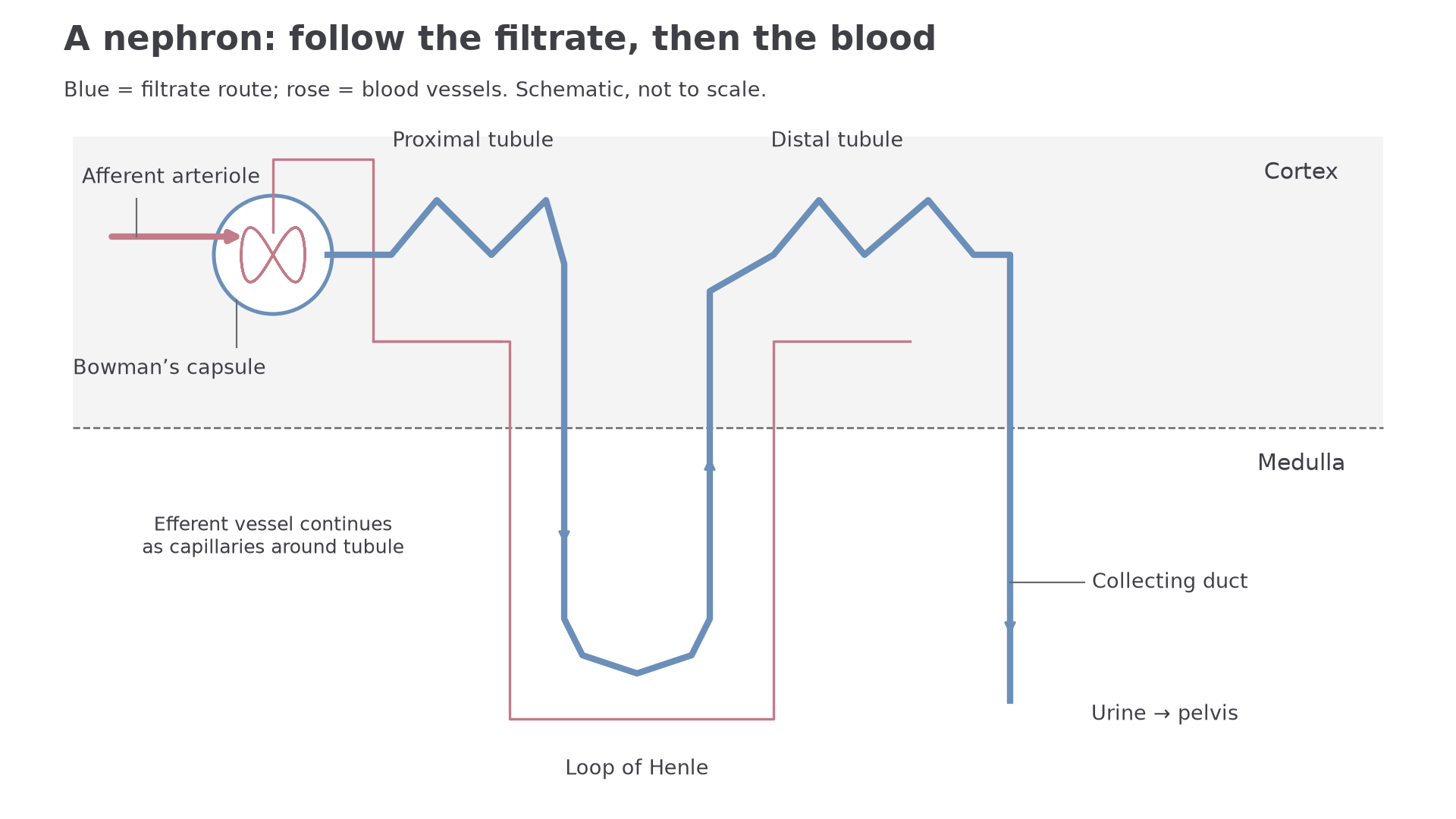
Fluid that enters Bowman's capsule is called filtrate. It flows through the proximal convoluted tubule, down and up the loop of Henle, through the distal convoluted tubule and into a collecting duct. The collecting ducts carry urine towards the renal pelvis.
The efferent arteriole forms capillaries around the tubule, including peritubular capillaries and the vasa recta. This close association is essential: useful substances move from the tubule fluid back into the blood over short distances.
When describing nephron action, keep two routes separate: blood flows through vessels, while filtrate flows through the tubule. Mixing those routes is a common way to lose marks.
The names are easier to remember if you link them to the process. The glomerulus filters. The proximal convoluted tubule reabsorbs most useful molecules. The loop of Henle helps create conditions for water reabsorption. The distal convoluted tubule and collecting duct make final adjustments and carry the remaining fluid onwards as urine.
Ultrafiltration
Ultrafiltration is the pressure-driven filtration of blood at the renal corpuscle. The word "ultra" matters because the filtration barrier is very fine: small molecules can pass into Bowman's capsule, while blood cells and most plasma proteins remain in the blood.
The pressure is high because blood arrives in the glomerulus through the afferent arteriole and leaves through the narrower efferent arteriole. Resistance to outflow helps maintain high hydrostatic pressure in the glomerular capillaries. Do not describe blood accumulating indefinitely: at steady state, inflow balances outflow plus filtered fluid. The pressure forces water and small solutes out of the blood plasma and across the filtration barrier.
Substances usually small enough to enter the filtrate include:
| Substance | In filtrate after ultrafiltration? | Reason |
|---|---|---|
| water | yes | small molecule |
| glucose | yes | small soluble molecule |
| amino acids | yes | small soluble molecules |
| urea | yes | small soluble waste molecule |
| inorganic ions | yes | small charged particles |
| red blood cells | no | too large and cellular |
| most plasma proteins | no | too large to pass through the filtration barrier |
This first step is not selective reabsorption. At ultrafiltration, useful small molecules such as glucose enter the filtrate along with waste molecules such as urea. The kidney recovers useful substances later.
Ultrafiltration separates by pressure and size. It does not yet decide which small molecules are useful.
The filtration barrier has three layers: fenestrations (small gaps) in the glomerular capillary endothelium, the basement membrane, and filtration slits between the foot processes of podocytes in Bowman's capsule. The basement membrane restricts large proteins; together the layers retain blood cells while water and small solutes pass. You do not need specialist medical pathology, but you do need the principle: the barrier lets small solutes and water through while retaining cells and large proteins in the blood.
Continue this lesson.
You’re previewing NovaLearn without an account. Create a free account to read the rest.
Create free account