5.1.4(c)(i)-(ii) - Pancreas histology and endocrine tissue
The pancreas is useful in hormonal communication because its endocrine tissue releases hormones into the blood. In this lesson you will learn how that endocrine tissue appears in stained pancreas sections, how it differs from the surrounding exocrine tissue, and how to make accurate observations and biological drawings from microscope slides. The focus is histology and drawing: detailed blood-glucose feedback belongs in the next lesson.
Pancreas As Mixed Tissue
Histology is the study of tissues using a microscope. A stained section of pancreas does not show one uniform tissue; it shows two main tissue roles packed together in the same organ.
Endocrine tissue
Endocrine tissue contains cells that secrete hormones directly into the blood.
That direct route into blood is what makes endocrine tissue important for hormonal communication.
Exocrine tissue
Exocrine tissue secretes substances into ducts, which carry the secretion to another surface or cavity.
Most of the pancreas is exocrine tissue. It is arranged into many small secretory units called acini. The acinar cells produce digestive secretions, and these secretions pass into ducts. You do not need the details of pancreatic digestive enzymes here; the key histology idea is that exocrine tissue is duct-linked.
The endocrine tissue occurs in clusters called islets of Langerhans, or pancreatic islets. These clusters contain hormone-secreting cells. At this level, the most important examples are beta cells, which secrete insulin, and alpha cells, which secrete glucagon. This lesson only uses those names to identify endocrine tissue; the control of insulin and glucagon secretion is taught later.
The structure-function link is direct: endocrine islets need access to blood because hormones are carried in the blood to target tissues. Exocrine acini need ducts because their secretions are not released directly into the bloodstream.
Recognising Islets In Stained Sections
In a typical stained light-microscope section, the endocrine islets usually appear as paler-staining rounded or irregular clusters among darker exocrine acini. The contrast is not because islets are empty spaces; it is because the endocrine cells are arranged differently and take up the stain differently from the surrounding acinar tissue.
[DIAGRAM: pancreas_histology_map: Lesson 107: Pancreas histology and endocrine tissue - diagram 01; asset_slug: 107_m05_1_4_pancreas_histology_and_endocrine_tissue__diagram_01; recommended_method: drawn_biology; description: 16:9 deterministic line diagram of a stained pancreas section, showing a pale endocrine islet among exocrine acini, a duct branch, a blood vessel, and biological drawing conventions.]
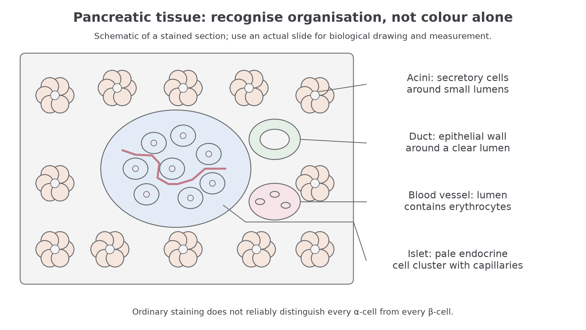
Use a consistent recognition route:
| Feature | How it may appear in a stained section | Why it matters |
|---|---|---|
| Islet of Langerhans | Paler cluster of many cells, often rounded or irregular | Endocrine tissue; hormone release into blood. |
| Exocrine acini | More numerous darker-staining secretory units | Exocrine tissue; secretions pass into ducts. |
| Duct branch | Tube-like structure with a clear lumen | Carries exocrine secretions away. |
| Blood vessel | Lumen that may contain red blood cells | Route for endocrine hormones to enter circulation. |
Do not expect a real section to look as neat as a textbook diagram. Tissue may be cut at an angle, staining may vary, and not every field of view will contain a perfect islet, duct and vessel together. The skill is to use the pattern: pale endocrine clusters embedded in a larger field of exocrine tissue.
A duct has a continuous epithelial lining around its lumen. A blood vessel may contain erythrocytes; a wider vessel containing many erythrocytes side by side should not automatically be called a capillary. A true capillary is narrow enough for red blood cells to pass largely in single file. Colour alone is insufficient: use cell arrangement, lumen and contents together.
Endocrine And Exocrine Contrast
The pancreas is sometimes described as having both endocrine and exocrine functions. In histology, that statement becomes visible as two different tissue organisations.
Continue this lesson.
You’re previewing NovaLearn without an account. Create a free account to read the rest.
Create free account