5.1.2(d) - Control of blood water potential by ADH
Your cells work properly only if the water potential of the blood is kept close to a set point. This lesson follows one homeostatic control loop: how osmoreceptors in the hypothalamus detect a change, how the posterior pituitary releases ADH, and how ADH changes the walls of the collecting ducts so urine volume changes.
Blood Water Potential
Water potential describes the tendency of water molecules to move. In this lesson, the important comparison is between the water potential of the blood and the set point that the body is trying to maintain.
If a person loses water through sweating, or takes in a lot of solute without enough water, the blood water potential falls. The blood becomes more concentrated: its osmolality (the number of dissolved particles per kilogram of solvent) and osmotic pressure increase. Osmolality and osmotic pressure are related but have different units; mOsm kg⁻¹ measures osmolality, not pressure. If a person drinks a large volume of water, blood water potential rises and the blood becomes less concentrated.
Osmoregulation
Osmoregulation is the homeostatic control of the water potential of body fluids, including blood plasma.
The kidney can adjust how much water remains in the urine. The part of the nephron controlled in this lesson is the collecting duct. Filtrate flows down the collecting duct towards the pelvis of the kidney. Water can leave the filtrate by osmosis if the collecting duct wall is permeable to water and the surrounding medulla/tissue fluid has a lower water potential than the filtrate.
That medullary water potential gradient is a prerequisite from kidney function, but it is not the focus here. This lesson is about how the hormone ADH changes the permeability of the collecting duct wall, so more or less water is reabsorbed into the blood.
Use "blood water potential" or "osmotic pressure" precisely. Do not replace the whole explanation with vague phrases such as "the blood is too watery" or "ADH adds water".
Detecting Low Water Potential
When blood water potential falls, water moves out of osmoreceptor cells in the hypothalamus by osmosis. The cells shrink slightly. That change affects neurosecretory cells in the hypothalamus, leading to the release of antidiuretic hormone, or ADH.
Osmoreceptor
An osmoreceptor is a sensory receptor that detects changes in the water potential or osmotic pressure of the blood.
ADH is made by neurosecretory cells in the hypothalamus. It then travels down the axons of these cells to the posterior pituitary gland, where it is stored in vesicles. When blood water potential is too low, impulses in the neurosecretory cells cause ADH to be released by exocytosis from the posterior pituitary into the blood.
[DIAGRAM: adh_feedback_control_loop: Lesson 099: Control of blood water potential by ADH - diagram 01; asset_slug: 099_m05_1_2_control_of_blood_water_potential_by_adh__diagram_01; recommended_method: drawn_biology; description: A clean negative feedback flow diagram showing low blood water potential stimulating hypothalamic osmoreceptors, ADH release from the posterior pituitary, increased collecting duct permeability, more water reabsorption, concentrated urine, and return towards normal blood water potential.]
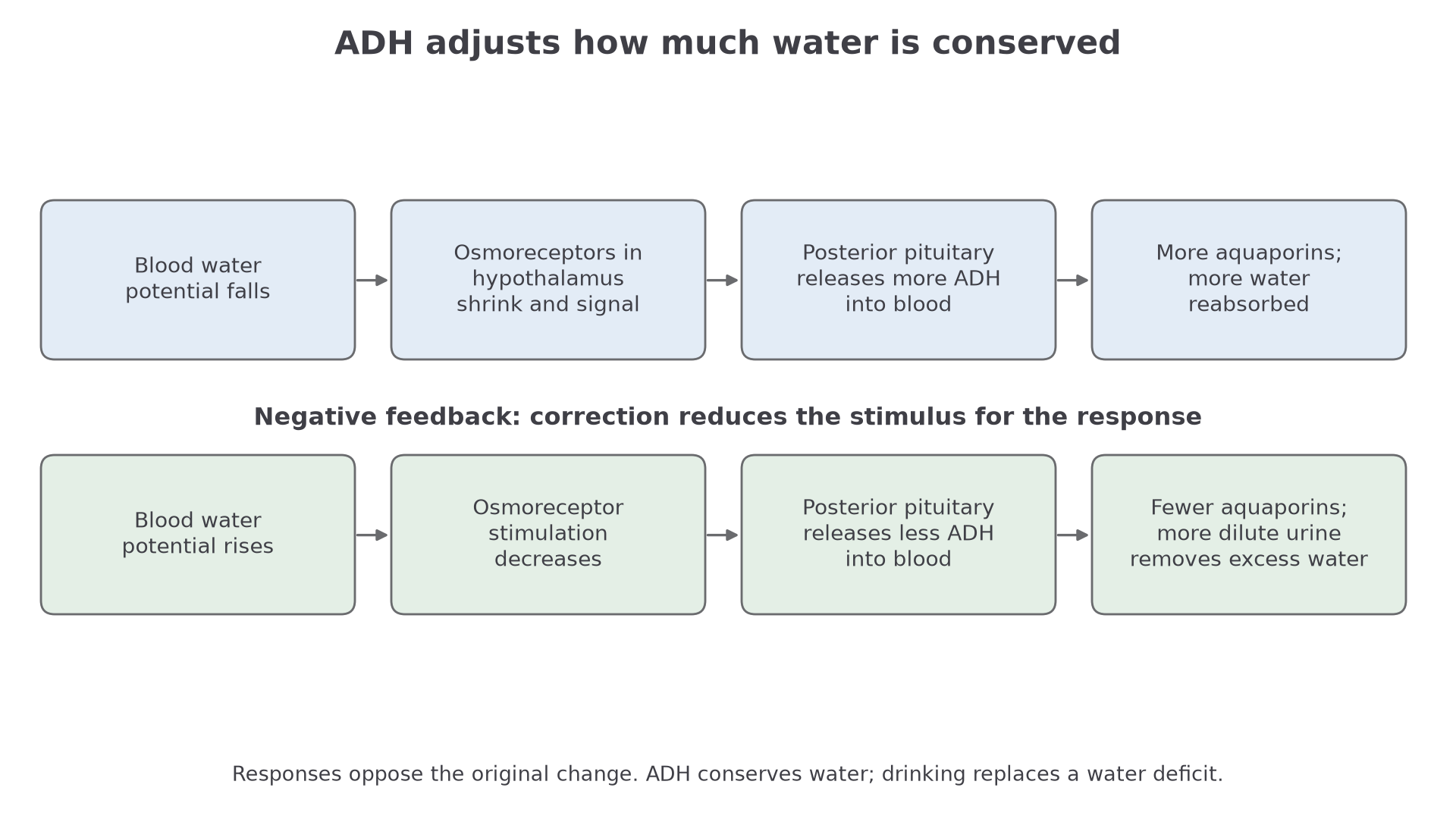
There are two distinct receptor roles: osmoreceptors detect the blood change, while ADH receptors on kidney cells detect the hormone. Follow the route between them:
| Stage | Biological sequence | Common error to avoid |
|---|---|---|
| detection | osmoreceptors in the hypothalamus detect low blood water potential | saying the kidney detects it directly |
| hormone source | ADH is made in the hypothalamus | saying ADH is made by the adrenal gland |
| release site | ADH is released from the posterior pituitary gland | saying anterior pituitary |
| transport | ADH travels in the blood to the kidney | saying ADH travels down the nephron |
ADH Action On Collecting Ducts
ADH is carried in the blood to the kidney. Its target cells are in the walls of the collecting ducts. These cells have receptors that can bind ADH, so they are target cells for the hormone.
When ADH binds to receptors on collecting duct cells, it triggers a signalling pathway inside the cell. The important outcome is that vesicles containing aquaporins move to and fuse with the cell surface membrane facing the filtrate.
Continue this lesson.
You’re previewing NovaLearn without an account. Create a free account to read the rest.
Create free account