2.1.6(c)-(d) - Mitosis stages and plant tissue microscopy
Mitosis is the part of the cell cycle where the copied genetic material is separated into two nuclei. In this lesson you will learn the visible stages of mitosis, the exact structures whose changes you must track, and how stained plant tissue can be used as evidence for those stages. The practical thread matters because microscope observations are only useful when you can describe, draw and interpret what is actually visible.
Mitosis As Nuclear Division
Mitosis is nuclear division. It separates copied chromosomes so that two new nuclei receive the same genetic information. The division of the cytoplasm, called cytokinesis, can begin during late mitosis and completes daughter-cell separation; it is closely linked in timing, but it is not the same process.
Before mitosis begins, DNA has already been copied during the previous interphase. Each copied chromosome consists of two identical sister chromatids joined at a centromere. A chromosome is easiest to see under a light microscope when it is condensed: long, diffuse chromatin becomes shorter, thicker, darker-staining chromosome material.
Sister Chromatids
The two identical copies of a replicated chromosome, joined together at the centromere until they separate during anaphase.
The centromere is the named region that makes that joined pair useful during mitosis, because it is where separation is organised.
Centromere
The region of a chromosome where sister chromatids are held together and where spindle fibres attach during mitosis.
The structures to follow are the nuclear envelope, chromosomes, chromatids, centromeres, centrioles, spindle fibres and cell membrane. Centrioles are spindle-organising structures usually shown in animal-cell mitosis diagrams; they are part of the OCR structure list, but they are not a feature you should expect to identify in a plant root-tip squash. In plant tissue microscopy, the clearest evidence is usually the pattern of stained chromatin or chromosomes inside cell outlines.
Prophase And Metaphase
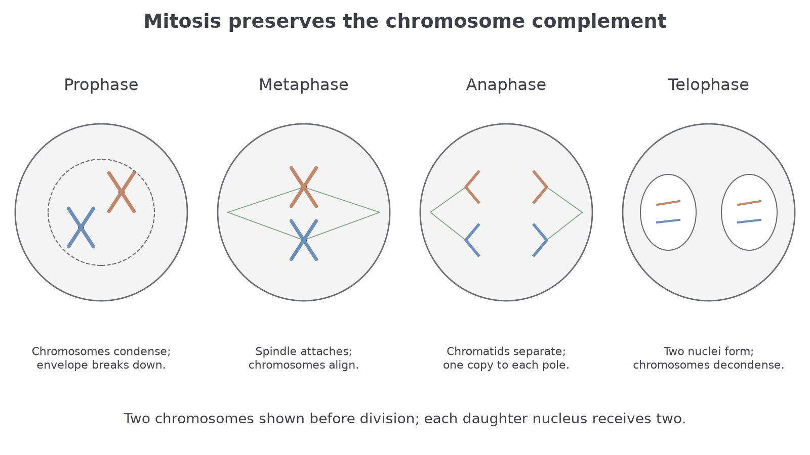
Prophase is the stage where the nucleus stops looking like a quiet, diffuse area and starts showing mitotic organisation. The chromosomes condense, so each replicated chromosome can be seen as two sister chromatids joined at a centromere. The nuclear envelope breaks down, allowing spindle fibres to interact with the chromosomes. In many diagrams, centrioles move to opposite poles and spindle fibres begin to form between them.
Metaphase is the stage where the chromosomes are arranged at the cell equator. The equator is the middle of the spindle, not a physical line drawn inside the cell. Spindle fibres attach to the chromosomes at the centromere region, so each sister chromatid is connected towards an opposite pole. This careful alignment matters for the visible sequence: a metaphase cell normally shows condensed chromosomes in a single central line or plate.
Do not describe metaphase as "chromatids separating". If chromatids are being pulled apart, the cell has moved into anaphase. A concise stage description should combine the visible arrangement with the named structure causing it: chromosomes align at the equator and spindle fibres attach at the centromeres.
When identifying a stage from a described microscope image, use visible evidence. "Chromosomes are lined up at the equator" is stronger than just naming the stage.
Now try turning the stage description into creditworthy wording.
The nucleolus also disappears during prophase. In an animal cell, a centrosome containing centrioles helps organise each spindle pole. Higher plant cells assemble a spindle without centrioles, so the same chromosome movements occur without the animal-cell pole structures.
[DIAGRAM: mitosis_stages: Chromosome behaviour across mitosis; asset_slug: 046_m02_1_6_mitosis_stages_and_plant_tissue_microscopy__diagram_01; recommended_method: drawn_biology; description: A simplified animal cell with a constant two-chromosome complement shows condensation, metaphase alignment, chromatid separation and two restored nuclei.]
Anaphase And Telophase
Anaphase begins when the centromeres divide. The sister chromatids separate and are pulled towards opposite poles by spindle fibres. Once separated, each chromatid is now functioning as a daughter chromosome. In a micrograph, anaphase is usually recognised by two groups of distinct, often V-shaped chromosomes moving apart.
Telophase is the reversal of much of prophase. The chromosomes have reached the poles and begin to decondense, becoming less individually distinct. New nuclear envelopes form around each group of chromosomes, so the cell starts to show two nuclei. Spindle fibres break down as the nuclear division finishes.
The cell membrane is involved in cytokinesis, which can overlap late mitosis and completes cell separation. In animal cells, the cell surface membrane constricts to divide the cytoplasm. In plant cells, a cell plate forms between the new nuclei and develops into the new separating wall and membranes. For this lesson, the important boundary is the distinction: mitosis divides the nucleus; cytokinesis completes cell division.
Anaphase separates sister chromatids. Telophase rebuilds nuclei around the separated chromosome groups.
The next check is deliberately about wording the visual evidence, because that is where many stage-identification errors happen.
Continue this lesson.
You’re previewing NovaLearn without an account. Create a free account to read the rest.
Create free account