5.1.5(l)(i)-(ii) - Mammalian muscle structure and contraction
Muscle contraction is a response mechanism: a nervous signal is converted into movement by protein filaments inside muscle fibres. In this lesson, you will compare the three mammalian muscle types, zoom into skeletal muscle structure, then follow the neuromuscular junction and sliding filament model. You will also learn how ATP is used and rapidly regenerated by creatine phosphate, and how stained skeletal muscle sections should be interpreted.
Muscle tissues compared
Mammals have three main muscle tissue types. The specification names them as skeletal, involuntary and cardiac muscle. Involuntary muscle here means smooth muscle: muscle in the walls of organs and blood vessels that is not under conscious control.
[DIAGRAM: muscle_type_comparison: Lesson 120: Mammalian muscle structure and contraction - diagram 01; asset_slug: 120_m05_1_5_mammalian_muscle_structure_and_contraction__diagram_01; recommended_method: drawn_biology; description: Three-panel deterministic comparison of skeletal, smooth/involuntary and cardiac muscle with structure cues including striations, nuclei, branching, intercalated discs and functional control.]
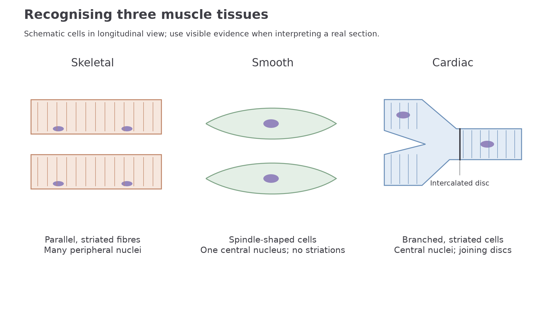
The first comparison is striation. Skeletal and cardiac muscle look striated because their actin and myosin filaments are arranged in repeating sarcomeres. Smooth muscle does not show striations because its contractile proteins are not arranged into the same regular banded pattern.
The second comparison is cell shape and nuclei. Skeletal muscle fibres are long cylindrical cells formed by fusion of many cells, so each fibre has many nuclei, usually near the edge. Smooth muscle cells are spindle-shaped with one central nucleus. Cardiac muscle cells are shorter and branched, usually with one central nucleus, and are joined by intercalated discs.
The third comparison is function. Skeletal muscle is mostly voluntary and produces movement of the skeleton. Smooth muscle contracts slowly and steadily in places such as the gut and blood vessel walls. Cardiac muscle is involuntary, striated and specialised for rhythmic contraction of the heart.
| Feature | Skeletal muscle | Involuntary muscle (smooth) | Cardiac muscle |
|---|---|---|---|
| Striations | Present | Absent | Present |
| Cell or fibre shape | Long cylindrical fibres | Spindle-shaped cells | Short branched cells |
| Nuclei | Many, usually peripheral | One central nucleus | Usually one central nucleus |
| Control | Mostly voluntary | Involuntary | Involuntary |
| Main role | Movement of skeleton | Slow movement/pressure in organs | Pumping blood |
Skeletal muscle hierarchy
Skeletal muscle structure is nested. A whole muscle contains bundles called fascicles. Each fascicle contains many muscle fibres. A muscle fibre is one long muscle cell, and it contains many myofibrils. Each myofibril is a chain of sarcomeres.
Sarcomere
The repeating contractile unit of a myofibril, running from one Z line to the next Z line, and containing overlapping actin and myosin filaments.
[DIAGRAM: skeletal_muscle_hierarchy: Lesson 120: Mammalian muscle structure and contraction - diagram 02; asset_slug: 120_m05_1_5_mammalian_muscle_structure_and_contraction__diagram_02; recommended_method: drawn_biology; description: Deterministic hierarchy diagram showing whole skeletal muscle, fascicle, muscle fibre, myofibril and one sarcomere as nested levels.]
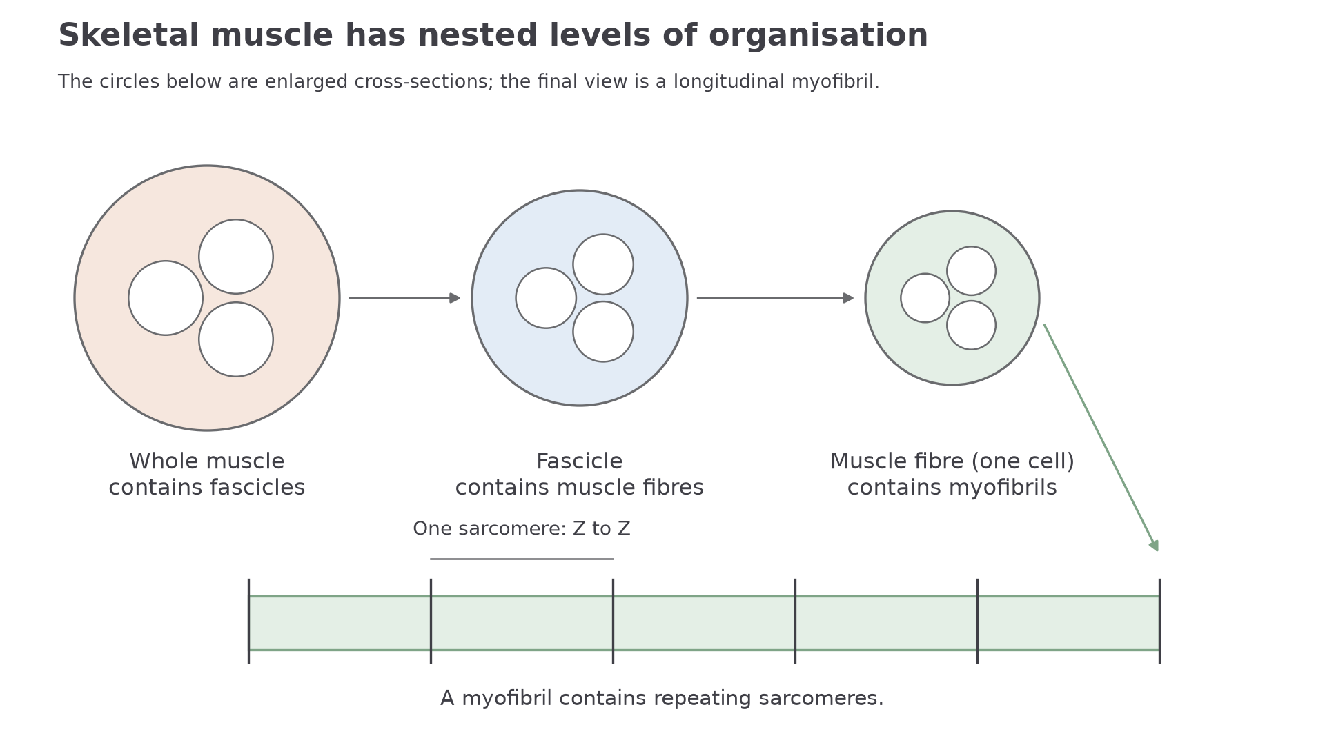
The key contractile proteins are actin and myosin. Actin forms the thin filaments. Myosin forms the thick filaments and has heads that can bind to actin. When many sarcomeres shorten at the same time, the myofibril shortens; when many myofibrils shorten, the muscle fibre contracts.
The striated appearance of skeletal muscle comes from the regular alignment of sarcomeres. Darker and lighter bands are not random stain differences. They reflect where thick and thin filaments overlap.
The route is: whole muscle -> fascicle -> muscle fibre -> myofibril -> sarcomere -> actin and myosin filaments.
Neuromuscular junction
Skeletal muscle fibres do not contract just because they contain actin and myosin. A contraction begins when a motor neurone stimulates the muscle fibre at a neuromuscular junction.
Neuromuscular junction
The synapse between a motor neurone and a skeletal muscle fibre, where acetylcholine transfers the signal from the neurone to the motor end plate.
[DIAGRAM: neuromuscular_junction_sequence: Lesson 120: Mammalian muscle structure and contraction - diagram 03; asset_slug: 120_m05_1_5_mammalian_muscle_structure_and_contraction__diagram_03; recommended_method: drawn_biology; description: Deterministic sequence diagram of a motor neurone terminal, synaptic cleft, ACh release, motor end plate depolarisation, T tubule signal and Ca²⁺ release from sarcoplasmic reticulum.]
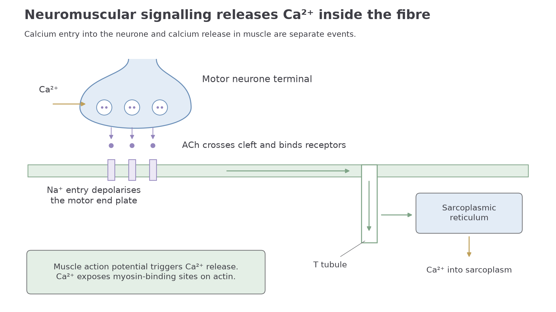
The sequence is:
- An action potential reaches the axon terminal of the motor neurone.
- Calcium ions enter the axon terminal.
- Vesicles release acetylcholine, often shortened to ACh, into the synaptic cleft.
- ACh diffuses across the cleft and binds to receptors on the motor end plate.
- Receptor channels open, allowing a net entry of Na⁺ into the muscle fibre. The motor end plate depolarises, starting an action potential in the muscle fibre membrane, the sarcolemma.
- The action potential spreads along the sarcolemma and down its inward folds, the T tubules. This triggers calcium-ion release from the sarcoplasmic reticulum into the sarcoplasm. The calcium entering the neurone terminal and the calcium released inside the muscle fibre are separate events.
- Calcium ions allow the sliding filament mechanism to begin.
After signalling, acetylcholinesterase breaks down ACh. This matters because it prevents continuous stimulation from one nerve impulse.
For neuromuscular junction questions, keep the direction clear: neurone action potential -> ACh release -> receptor binding on the muscle fibre -> muscle fibre depolarisation -> calcium ion release.
Use that sequence in the check below.
Continue this lesson.
You’re previewing NovaLearn without an account. Create a free account to read the rest.
Create free account