4.1.1(e)(i)-(ii) - Phagocytes, blood smears and phagocytosis
When a pathogen gets past the body's primary defences, some white blood cells respond by engulfing and digesting it. In this lesson you will learn the required structure and mode of action of phagocytes, then connect that biology to PAG1 blood-smear microscopy and biological drawing. The focus is phagocytes, especially neutrophils and antigen-presenting cells, not the later lymphocyte response.
Phagocytes and visible structure
A phagocyte is a white blood cell that can engulf particles, including pathogens, by phagocytosis. The word is useful because it describes a job, not just one cell type: a neutrophil is a phagocyte, and macrophages and dendritic cells are phagocytes that can also act as antigen-presenting cells.
Phagocyte
A white blood cell that engulfs and digests pathogens or other particles by phagocytosis.
The main phagocyte you can recognise in an ordinary blood smear is the neutrophil. A neutrophil has a flexible cell surface, a cytoplasm containing enzyme-rich granules and lysosomes, and a lobed nucleus. The flexible surface matters because the cell has to change shape and extend around a pathogen. The lysosomes matter because engulfing a pathogen is not enough; the pathogen must be broken down.
Neutrophil
A phagocytic white blood cell with a lobed nucleus, granular cytoplasm and enzyme-containing lysosomes, commonly seen in blood smears.
An antigen-presenting cell displays antigen fragments on its surface. In this lesson, the examples are antigen-presenting phagocytes that digest pathogen material before presenting it. Macrophages and dendritic cells are the clearest examples at this level. This display is the bridge from non-specific phagocytosis to the later specific immune response, but the details of B cells, T cells and clonal selection belong in the next lesson.
Antigen-presenting cell
A cell, such as a macrophage or dendritic cell, that presents antigen fragments on its surface after digesting a pathogen.
The key structure-function link is simple: a phagocyte is built for movement, recognition, engulfment and digestion. A lobed nucleus helps a neutrophil squeeze through small gaps between cells; a flexible membrane forms projections around a pathogen; lysosomes supply digestive enzymes.
Phagocytosis sequence
Phagocytosis involves more than engulfment. The pathogen must be recognised, enclosed inside a vesicle, digested, and in some cells its antigens may be presented.
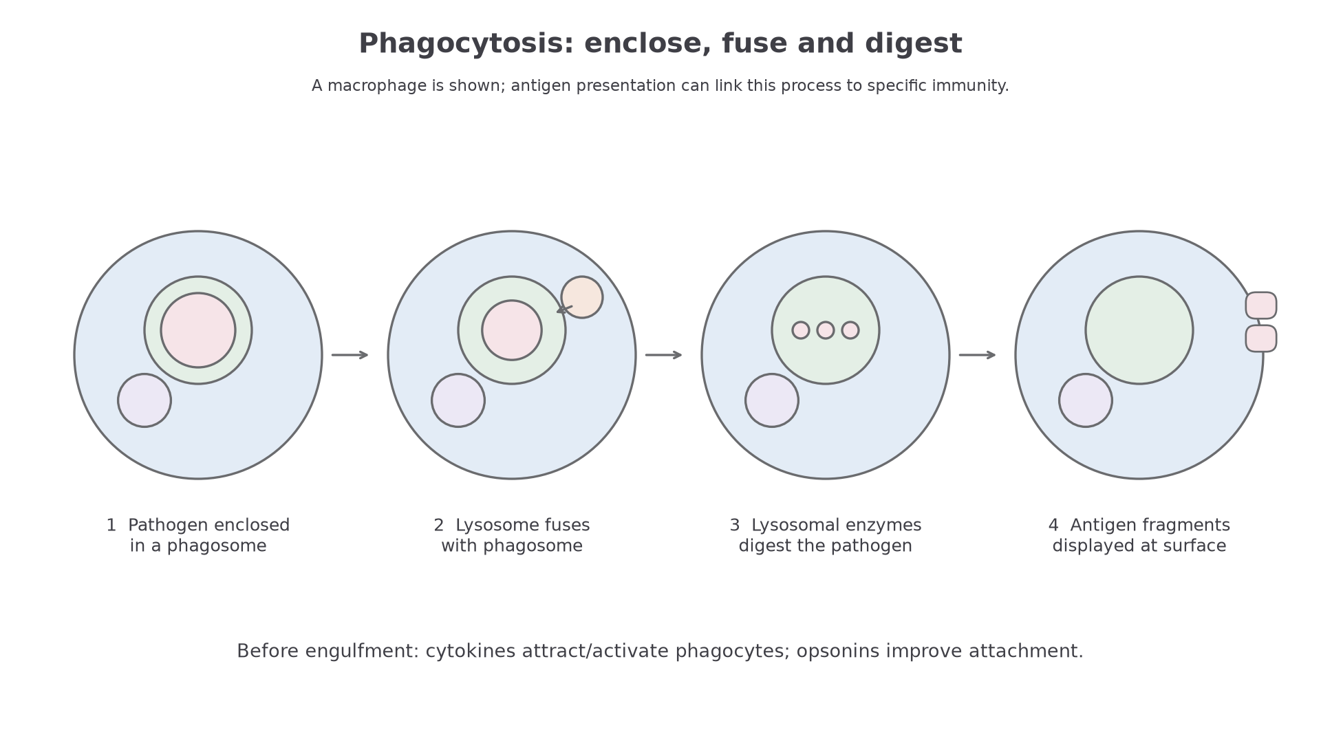
First, the phagocyte is attracted towards the site of infection by chemical signals. It then recognises molecules on the pathogen surface, or recognises molecules coating the pathogen. The phagocyte's membrane extends around the pathogen as projections called pseudopodia. When the membrane encloses the pathogen, a vesicle forms inside the phagocyte.
Phagosome
A vesicle inside a phagocyte that contains an engulfed pathogen or particle.
The phagosome then fuses with lysosomes. Lysosomes are membrane-bound vesicles containing digestive enzymes. After fusion, the enzymes break down the pathogen into smaller molecules and antigen fragments. You may see the fused digestive vesicle called a phagolysosome, but the required idea is that the phagosome fuses with lysosomes and lysosomal enzymes digest the pathogen.
The sequence is recognition, engulfment, phagosome formation, lysosome fusion, digestion, and sometimes antigen presentation.
[DIAGRAM: asset_slug: 073_m04_1_1_phagocytes_blood_smears_and_phagocytosis__diagram_01; description: Stages of engulfment, phagosome formation and lysosome fusion.]
Cytokines, opsonins and APCs
Two helper ideas make phagocytosis more efficient: cytokines help cells communicate, and opsonins make pathogens easier for phagocytes to recognise.
Cytokines are chemical signalling molecules released by cells, including damaged cells and immune cells. In this lesson, their important role is to attract or activate phagocytes and coordinate their movement towards infection. Their concentration can guide phagocytes towards the infection. You do not need to learn named cytokines here.
Continue this lesson.
You’re previewing NovaLearn without an account. Create a free account to read the rest.
Create free account