3.1.2(g)-(h) - Heart conduction and ECG interpretation
The heart does not wait for a nerve impulse before every beat. Cardiac muscle is myogenic, so it can initiate its own electrical excitation, then coordinate that excitation so the atria and ventricles contract in the right order. In this lesson you will connect the named conduction tissues to the pattern on an electrocardiogram, or ECG, and use trace data to recognise normal rhythm, tachycardia, bradycardia, fibrillation and ectopic heartbeat.
Myogenic Heart Action
Cardiac muscle is described as myogenic because the stimulus for contraction originates within the muscle itself. This does not mean every cardiac muscle cell fires randomly. A normal heartbeat is coordinated by specialised regions of cardiac tissue that initiate and conduct a wave of excitation.
Myogenic
Myogenic heart action means that cardiac muscle initiates its own contraction rather than needing an external nerve impulse to start each beat.
The key starting point is the sino-atrial node, usually shortened to SAN. The SAN is a small region of specialised tissue in the wall of the right atrium. It acts as the heart's natural pacemaker because it initiates the wave of excitation that spreads across the atrial walls.
As the wave of excitation spreads across the atria, the atria contract. This contraction helps push blood into the ventricles before the ventricles contract. The order matters: if atria and ventricles contracted at the same time, ventricular filling would be less effective.
Coordinating The Beat
After excitation spreads across the atria, it reaches the atrio-ventricular node, or AVN. The AVN is important because it delays the wave of excitation before passing it on. That short delay gives the atria time to finish contracting and empty more blood into the ventricles before ventricular systole begins.
A band of non-conducting tissue separates the atria from the ventricles, preventing direct spread of excitation into the ventricular muscle. The conducting route passes from the AVN into the bundle of His in the septum, then through purkyne tissue (Purkinje fibres) to the apex and up the ventricular walls.
[DIAGRAM: cardiac_conduction_pathway: Lesson 063: Heart conduction and ECG interpretation - diagram 01; asset_slug: 063_m03_1_2_heart_conduction_and_ecg_interpretation__diagram_01; recommended_method: drawn_biology; description: deterministic grey line diagram of the myogenic conduction pathway showing SAN, AVN, purkyne tissue, apex and spread of excitation through the ventricle walls.]
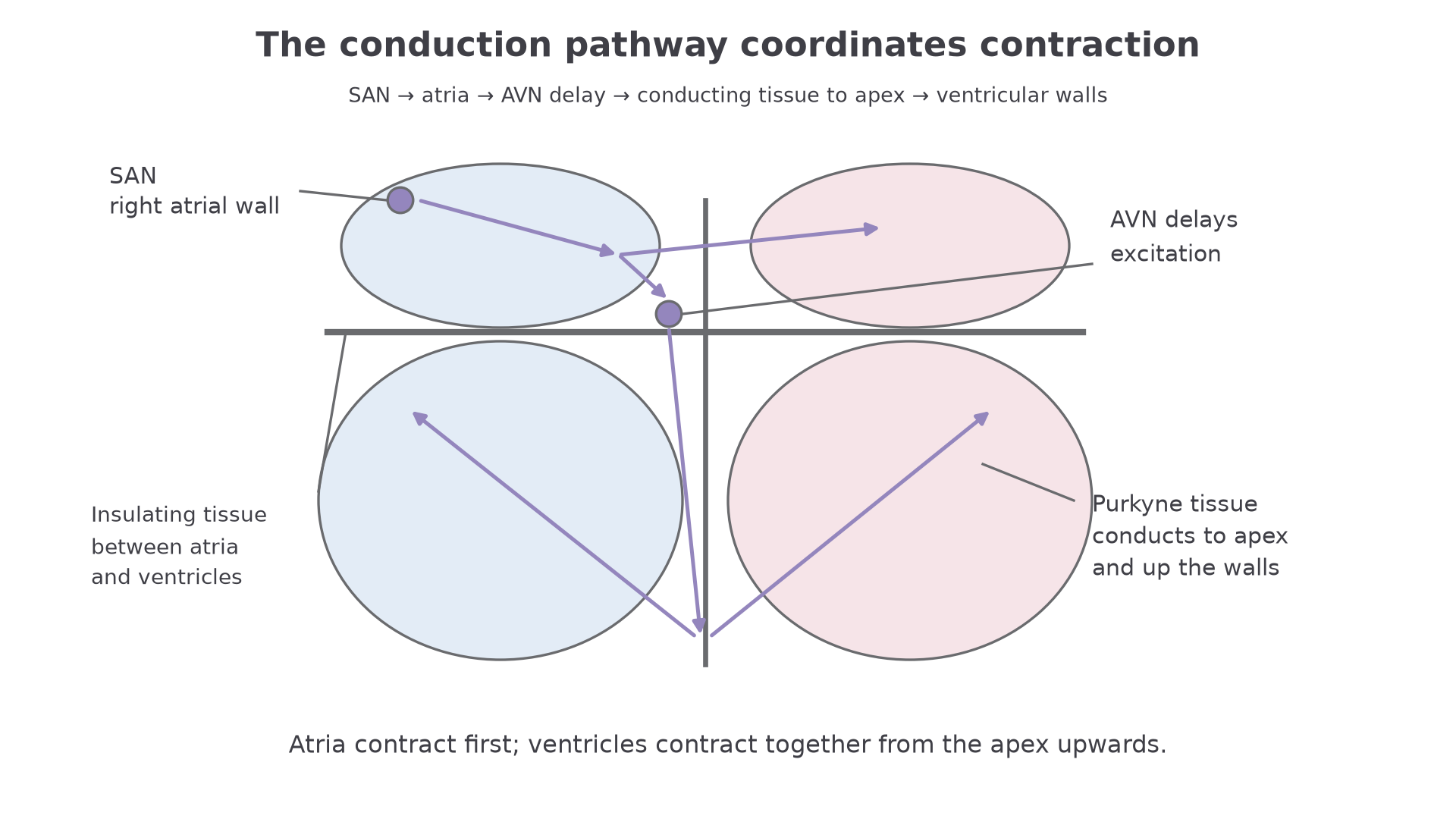
This route coordinates the mechanics of pumping. The ventricles are excited from the apex upwards, so contraction starts near the bottom of the ventricles and pushes blood up towards the exits: the pulmonary artery and aorta. That makes the sequence a structure-function story, not a list of names.
The complete sequence is:
| Stage | Role in coordination |
|---|---|
| SAN initiates excitation | starts the wave of excitation in the right atrium |
| Excitation spreads across atria | causes atrial contraction |
| AVN delays excitation | allows atria to empty before ventricles contract |
| Purkyne tissue conducts excitation | carries excitation to the apex and ventricle walls |
| Ventricles contract from apex upwards | helps force blood out through the arteries |
Normal ECG Trace
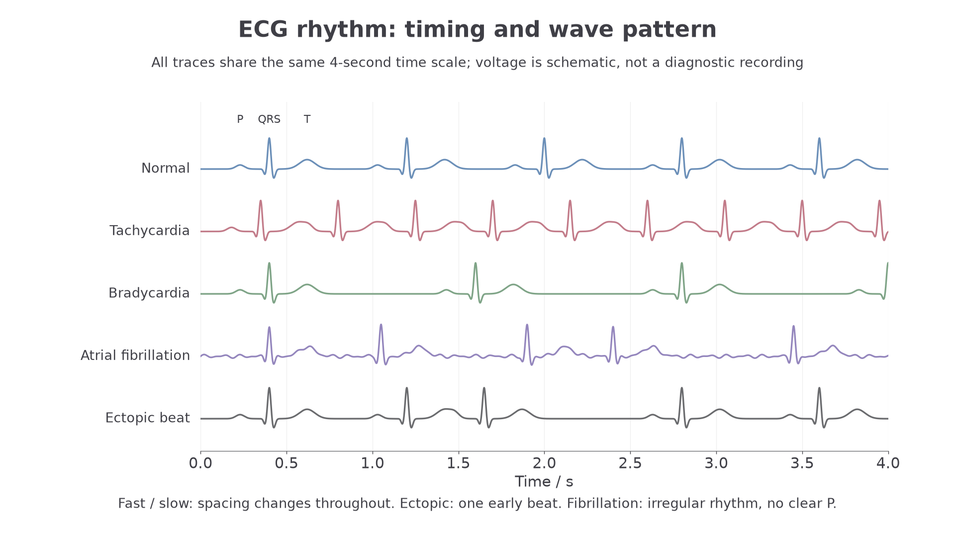
An electrocardiogram, or ECG, records electrical activity of the heart detected at the body surface. It is not a direct recording of blood pressure, heart sounds or the force of contraction. It gives evidence about when different regions of the heart are electrically excited and when the ventricles recover ready for the next beat.
In a simple normal ECG trace, one heartbeat is usually described using three main features:
| ECG feature | What it shows | Linked heart activity |
|---|---|---|
| P wave | atrial depolarisation | atria are electrically excited, leading to atrial systole |
| QRS complex | ventricular depolarisation | ventricles are electrically excited, leading to ventricular systole |
| T wave | ventricular repolarisation | ventricles recover electrically before the next beat |
The word depolarisation means electrical excitation of the muscle membrane. Repolarisation means the membrane returns towards its resting electrical state. For this lesson, those meanings are enough; detailed ion movements in cardiac cells are outside the specification point.
[DIAGRAM: ecg_trace_interpretation: Lesson 063: Heart conduction and ECG interpretation - diagram 02; asset_slug: 063_m03_1_2_heart_conduction_and_ecg_interpretation__diagram_02; recommended_method: drawn_biology; description: deterministic ECG interpretation panel showing normal rhythm, tachycardia, bradycardia, fibrillation and ectopic heartbeat with trace-spacing cues and a heart-rate formula.]
Continue this lesson.
You’re previewing NovaLearn without an account. Create a free account to read the rest.
Create free account