3.1.2(e)(i)-(ii) - Mammalian heart structure and dissection
The mammalian heart is two muscular pumps joined in one organ: the right side sends blood to the lungs, and the left side sends blood to the rest of the body. In this lesson you will learn the external and internal structures that make that separation possible, then use the same structures to plan a safe dissection, examination and biological drawing. The aim is not to memorise a polished textbook picture, but to recognise real features in a mammalian heart and explain what each one shows about function.
External Orientation
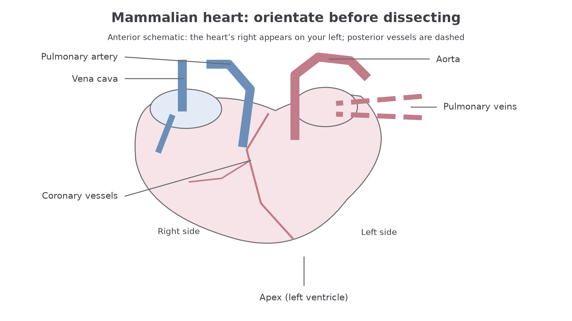
Before cutting a heart, orient it. A real sheep, pig or ox heart may be slippery, misshapen after storage, partly covered in fat and have vessels cut short, so orientation has to use several clues rather than one perfect photograph.
The broad base is the top of the heart where the large blood vessels enter or leave. The pointed apex lies lower down and is formed mainly by the left ventricle. On the anterior surface, the pulmonary artery and aorta leave from the top of the ventricles, and coronary vessels may be visible on the surface of the heart muscle. Coronary arteries supply oxygen and nutrients to the heart muscle itself; blood inside the chambers cannot supply all of the thick myocardium by diffusion.
The right and left sides are named as the heart's own right and left, not your right and left as you look at a diagram. In many diagrams, the right side of the heart appears on the left of the page because you are facing the front of the heart.
Use the vessels to map the two circuits:
| Structure seen externally | Main connection |
|---|---|
| Vena cava | brings deoxygenated blood from the body to the right atrium |
| Pulmonary artery | carries deoxygenated blood from the right ventricle to the lungs |
| Pulmonary veins | bring oxygenated blood from the lungs to the left atrium |
| Aorta | carries oxygenated blood from the left ventricle to the body |
Define arteries and veins by direction of blood flow, not by oxygen content. The pulmonary artery carries blood away from the heart even though it is deoxygenated, and the pulmonary veins carry blood towards the heart even though they are oxygenated.
[DIAGRAM: 061_m03_1_2_mammalian_heart_structure_and_dissection__diagram_01: Anterior heart orientation with major vessels, coronary vessels and left ventricular apex; asset_slug: 061_m03_1_2_mammalian_heart_structure_and_dissection__diagram_01; recommended_method: drawn_biology; description: Anterior heart orientation with major vessels, coronary vessels and left ventricular apex.]
Internal Structure
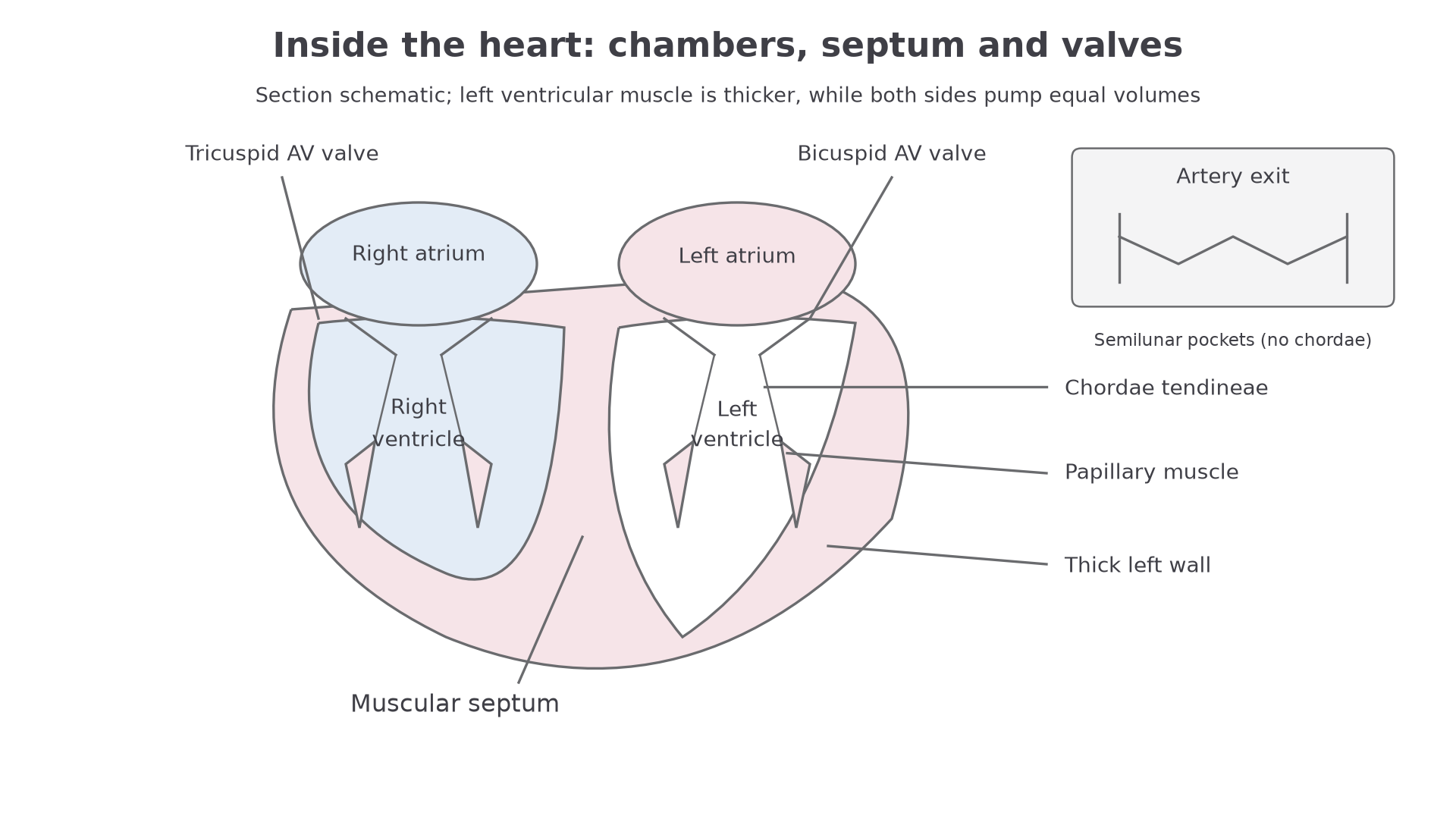
Inside the mammalian heart there are four chambers: two atria and two ventricles. Atria receive blood returning to the heart. Ventricles pump blood out of the heart. The right atrium and right ventricle form the right pump; the left atrium and left ventricle form the left pump.
The septum is the muscular wall separating the right and left sides. It prevents oxygenated and deoxygenated blood from mixing. This matters because the left side can then send oxygen-rich blood to body tissues, while the right side sends deoxygenated blood to the lungs.
The ventricles have thicker walls than the atria because ventricles pump blood out of the heart. The left ventricle has the thickest wall because it must generate enough pressure to send blood around the systemic circulation. The right ventricle has a thinner wall because it sends blood only to the lungs, a shorter route where excessive pressure would damage delicate exchange surfaces.
Valves keep blood moving in one direction. The atrioventricular valves lie between atria and ventricles: the tricuspid valve is on the right side and the bicuspid, or mitral, valve is on the left side. These valves are held by chordae tendineae attached to papillary muscles in the ventricle walls. The chordae tendineae stop the valve flaps from being forced back into the atria.
Semilunar valves lie at the exits from the ventricles: the pulmonary semilunar valve leads to the pulmonary artery and the aortic semilunar valve leads to the aorta. They are pocket-like valves at the vessel bases. You need their position and role in one-way flow here; detailed pressure changes during the cardiac cycle belong in the next lesson.
[DIAGRAM: 061_m03_1_2_mammalian_heart_structure_and_dissection__diagram_02: Connected heart section showing chambers, septum, ventricular muscle and AV valve support; semilunar valve inset; asset_slug: 061_m03_1_2_mammalian_heart_structure_and_dissection__diagram_02; recommended_method: drawn_biology; description: Connected heart section showing chambers, septum, ventricular muscle and AV valve support; semilunar valve inset.]
What Dissection Reveals
A dissection turns a diagram into evidence. When you examine a real heart, look for features that explain the diagram rather than trying to force the specimen to match a diagram perfectly.
Continue this lesson.
You’re previewing NovaLearn without an account. Create a free account to read the rest.
Create free account