2.1.6(i) - Cells, tissues, organs and organ systems
Multicellular organisms are not just large bags of separate cells. Their cells are organised into levels, and each level adds coordination: cells work together as tissues, tissues combine as organs, and organs cooperate in organ systems. In this lesson you will learn the hierarchy and the tissue examples named for this specification point: squamous and ciliated epithelia, cartilage, muscle, xylem and phloem.
Levels of organisation
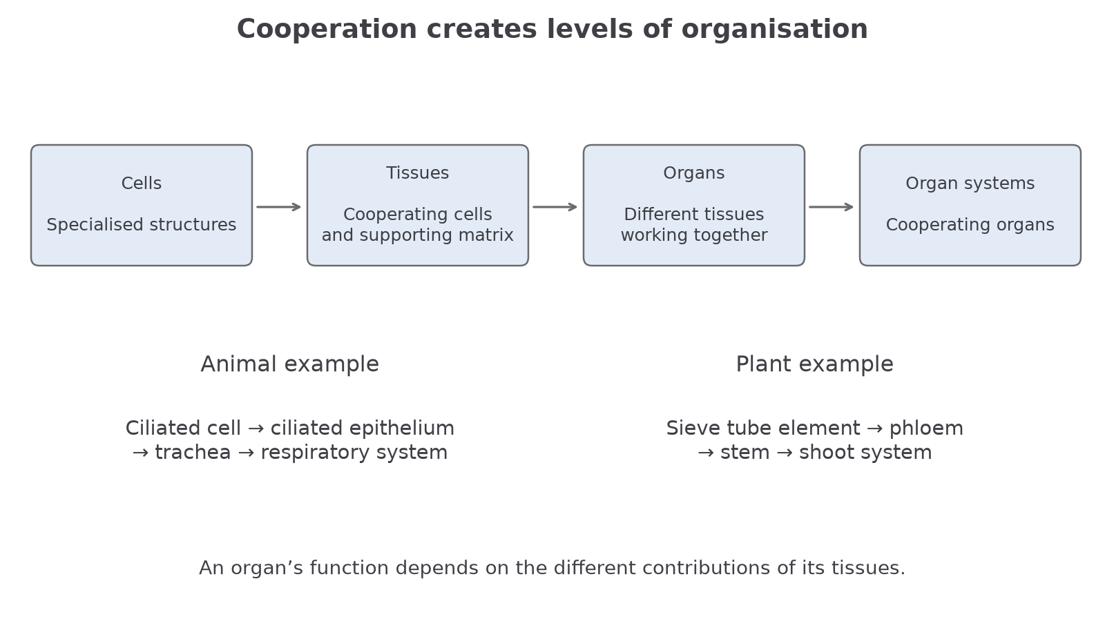
A specialised cell has features that help it carry out a particular role. A tissue is the next level up: it is a group of cells working together to perform a shared function. This definition should stay flexible enough for real biology, because some tissues include more than one cell type and some include extracellular material secreted by the cells.
Tissue
A group of cells, often with supporting extracellular material, working together to perform a particular function.
An organ is made from different tissues working together. For example, a plant leaf is an organ because it contains several tissues that cooperate in photosynthesis, gas exchange and transport. An organ system is a group of organs working together for a major function, such as the respiratory system in an animal or the shoot system in a plant.
Organ
A structure made from different tissues working together to perform one or more functions.
The next level up is about cooperation between whole organs rather than cooperation between tissues inside one organ.
Organ system
A group of organs working together to carry out a major function in an organism.
The key idea is not size alone. A tissue is not simply "a bigger cell"; it is a coordinated level of organisation.
[DIAGRAM: organisation_hierarchy_and_tissue_examples: Lesson 050: Cells, tissues, organs and organ systems - diagram 01; asset_slug: 050_m02_1_6_cells_tissues_organs_and_organ_systems__diagram_01; recommended_method: drawn_biology; description: A clean hierarchy from specialised cells to tissues, organs and organ systems, with the OCR-required animal and plant tissue examples.]
Animal tissue examples
An epithelium is a tissue that covers a surface or lines a space. The plural is epithelia. This specification point names two epithelial tissues: squamous epithelium and ciliated epithelium.
Squamous epithelium is made of thin, flattened cells. In exchange surfaces such as alveoli, a very thin squamous lining gives a short diffusion distance, so gases can move across quickly. The structure-function link is the important part: thin cells make a thin barrier.
Ciliated epithelium contains cells with cilia on the exposed surface. Cilia beat in a coordinated way, moving mucus or other material along the surface. In airways, mucus traps dust and microorganisms, and cilia help move the mucus away from delicate gas-exchange surfaces.
Cartilage is a connective tissue. It contains cells in a firm extracellular matrix, which makes it useful for support, cushioning and keeping some passages open. Muscle is a contractile tissue: its cells can shorten, so muscle tissue can produce movement. You do not need a detailed account of every muscle type in this lesson; the tissue-level function is contraction.
| Tissue example | Key tissue idea | Structure-function link |
|---|---|---|
| Squamous epithelium | Thin epithelial lining | Short diffusion distance |
| Ciliated epithelium | Epithelial lining with cilia | Moves mucus or other material along a surface |
| Cartilage | Connective tissue with firm matrix | Support and cushioning |
| Muscle | Contractile tissue | Movement by contraction |
If a question asks for a tissue, answer at tissue level. For example, "ciliated epithelium" is the tissue; "ciliated cell" is a cell type within that tissue.
The check below asks you to keep the two epithelial tissues separate by linking each structure to its function.
Cartilage cells, called chondrocytes, secrete and occupy spaces in an extracellular matrix containing fibres. The matrix resists compression, so cartilage can cushion a joint and support an airway even though most of its volume is not cells. Muscle tissue contains elongated cells with contractile proteins and mitochondria that supply ATP; many cells contracting together exert force.
An epithelial sheet has closely joined cells on a basement membrane. In an airway, ciliated cells and mucus-secreting goblet cells cooperate within the lining. This shows why a tissue need not contain only one cell type.
Continue this lesson.
You’re previewing NovaLearn without an account. Create a free account to read the rest.
Create free account