5.1.2(b)(i)-(ii) - Liver structure, histology and functions
The liver is part of excretion because it removes or changes harmful metabolic products before they disrupt homeostasis. In this lesson you will link visible liver structure to microscopic histology, then use that structure to explain glycogen storage, detoxification and urea formation. You will also practise the microscope and biological drawing decisions OCR expects for stained liver sections.
Gross Structure And Blood Supply
The mammalian liver is a large, lobed organ with a very high blood supply. Its position in the circulation is the key to its functions: blood from the gut reaches the liver before returning fully to the rest of the body.
Three gross structures matter most here:
| Structure | What it carries | Why that helps the liver |
|---|---|---|
| Hepatic portal vein | Nutrient-rich blood from the digestive system | Hepatocytes can store or process absorbed substances before they circulate widely. |
| Hepatic artery | Oxygenated blood from the heart | Liver cells have enough oxygen for active metabolism. |
| Hepatic vein | Blood leaving the liver | Processed blood returns towards the heart. |
The main working cells of the liver are hepatocytes. They sit next to blood spaces, so substances can move between the blood and the cells quickly.
Hepatocyte
A liver cell that carries out metabolic functions such as glycogen storage, detoxification and urea formation.
The liver also contains bile ducts. Bile is useful as a structural clue in histology because bile duct branches occur with blood-vessel branches in portal triads. Detailed bile function is not the focus of this lesson.
Liver Lobule Histology
Under the microscope, liver tissue is organised into repeating units called lobules. A classic liver lobule is often drawn as roughly hexagonal. A central vein lies in the middle, while portal triads lie near the edges.
[DIAGRAM: liver_lobule_histology_map: Lesson 097: Liver structure, histology and functions - diagram 01; asset_slug: 097_m05_1_2_liver_structure_histology_and_functions__diagram_01; recommended_method: drawn_biology; description: Labelled 16:9 line diagram of a classic mammalian liver lobule, showing portal triads with hepatic portal vein branch, hepatic artery branch and bile duct branch, plus hepatocyte plates, sinusoids, central vein, blood-flow direction towards the central vein and bile-flow direction towards the bile duct.]
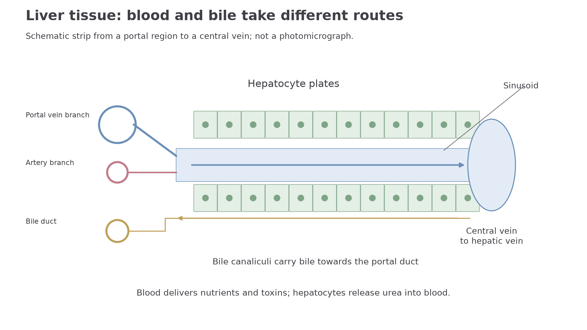
Each portal triad contains:
- a branch of the hepatic portal vein, usually the largest lumen;
- a branch of the hepatic artery, usually smaller with a thicker wall;
- a bile duct branch.
Blood from the hepatic portal vein and hepatic artery enters sinusoids. The portal supply is less oxygenated because it has already passed through respiring gut tissues; the arterial supply delivers oxygen at higher pressure. The thicker muscular wall of an arterial branch distinguishes it from the larger, thinner-walled portal venule. These are blood spaces between plates of hepatocytes. The blood flows through the sinusoids towards the central vein, giving hepatocytes direct access to dissolved nutrients, toxins and nitrogenous waste products.
Sinusoid
A blood-filled space in liver tissue that carries mixed blood past hepatocytes towards the central vein.
Blood runs from portal vessels through sinusoids to the central vein. Bile takes a different route: hepatocytes secrete it into tiny canaliculi between adjacent cells, which carry it towards bile ducts in the portal triads. A duct has a lining of epithelial cells; it is not another blood vessel. Central veins drain towards the hepatic vein.
Storage And Detoxification
The first specified function is storage of glycogen. After a meal, blood arriving from the gut may contain a high concentration of glucose. Hepatocytes can convert glucose into glycogen and store it. Glycogen is a compact, insoluble storage carbohydrate, so storing glucose in this form avoids making the cell contents too concentrated.
Later, glycogen can be broken back down to glucose when the body needs it. This lesson is about the liver role itself; detailed hormonal control of blood glucose is taught later.
The second specified function is detoxification. Detoxification means changing a harmful substance into a less harmful substance, or into a form that can be excreted more easily. Because blood from the gut passes through the liver, hepatocytes are well placed to metabolise ingested toxins, including some drugs and alcohol.
Continue this lesson.
You’re previewing NovaLearn without an account. Create a free account to read the rest.
Create free account