24.3 - Positrons and PET imaging
Explain how a beta-plus tracer leads to detectable gamma-ray pairs. Use mass–energy and momentum conservation, calculate annihilation photon energy, and distinguish a coincidence line from the concentration image reconstructed from many events.
A beta-plus tracer marks tissue uptake
Does detecting two PET photons tell you the exact point where a tracer nucleus decayed? No. The positron travels before annihilation, and one photon pair initially identifies a line of possible annihilation locations. The useful image emerges from many events.
Tracer
A tracer is a substance containing radioactive nuclei that is introduced into the body and absorbed by the tissue being studied.
The chemical substance determines how the tracer is distributed and taken up. Its radioactive nuclei provide a signal that can be detected from outside the body. Uptake need not be identical in all tissues, and not every nucleus is in the target tissue. PET, positron emission tomography, uses a tracer that decays by emission.
Recall the nuclear bookkeeping: a proton changes into a neutron, emitting a positron and an electron neutrino. Thus the daughter has one fewer proton, the same nucleon number, and atomic number one lower. In symbols,
This recalls the complete beta-plus equation; it is not a new supplied formula. The decay must be energetically possible. The positron is the electron's antiparticle, with the same rest mass and opposite electric charge.
The positron loses kinetic energy through interactions in the surrounding tissue and travels a finite distance before annihilation with an electron. The annihilation position is therefore generally displaced from the original decay position. This positron range contributes to the finite spatial resolution of PET; the scanner cannot remove that underlying physical displacement merely by making a sharper display.
Watch out
The scanner detects gamma-ray photons that escape from the body. It does not build the image by detecting positrons that have travelled from the tissue to external detectors.
PET primarily maps the distribution of tracer uptake and hence information about function. An X-ray or CT image principally maps attenuation and structure. Combining their information can relate tracer distribution to anatomy, but these are different measured quantities.
Annihilation conserves energy and momentum
When a positron interacts with an electron, the particle–antiparticle pair can annihilate. Their rest energy, together with any kinetic energy, is transferred to other forms of energy. In the PET model, two gamma-ray photons carry it away.
Mass alone is not conserved in this process: the electron and positron no longer remain as particles with their original rest masses. Total mass–energy and total momentum are conserved. Photons have zero rest mass, but they carry energy and momentum.
For the standard PET calculation, take the electron and positron to have negligible kinetic energy and negligible total momentum immediately before annihilation. The two photons then have equal energies and equal momentum magnitudes. To leave total momentum zero, they travel in opposite directions.
[DIAGRAM: asset_name: Positron range and annihilation; asset_slug: positron-annihilation; caption: The annihilation point is displaced from the decay and the photon momenta balance; description: A radioactive decay location is separated from a later electron-positron annihilation location by a short curved positron track. Two oppositely directed wavy photon arrows start at the annihilation location, with equal energy labels. The electron-positron pair is treated as nearly at rest immediately before annihilation. A note states that positions and distances are schematic and the photon symbols are representations of electromagnetic radiation.]
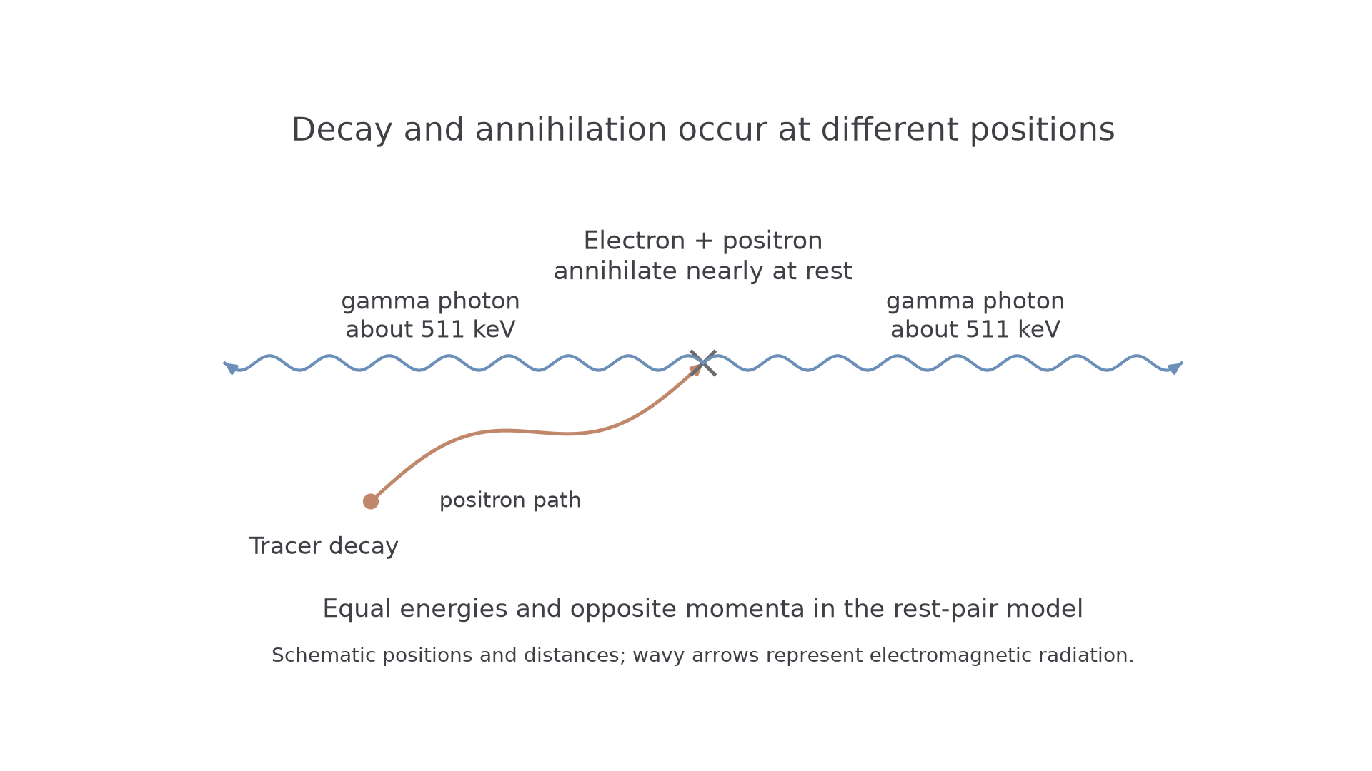
A single photon could carry away energy, but its nonzero momentum could not equal an initially zero total momentum. Two equal and opposite photon momenta solve both conservation requirements. The reason for zero initial total momentum is the stated near-rest approximation, not merely the fact that electron and positron have equal masses.
Model limit
The ideal PET picture uses a nearly stationary pair and two almost exactly opposite photons. Residual motion gives small departures from perfect opposition and equal energy. Do not apply the exact rest-pair result to an arbitrary fast-moving pair without checking its total momentum and energy.
The wavy arrows map photon propagation; photons are quanta of electromagnetic radiation, rather than tiny material balls. Gamma-ray photons can escape the body to be detected, although some are absorbed or scattered and do not provide a useful coincidence event.
Each rest-pair photon carries one electron rest energy
The pair has twice the rest energy of one electron. In the near-rest, two-photon model, each photon receives half that total. Forgetting the second particle or forgetting to divide between two photons gives the same factor-of-two trap in different disguises.
Rest-pair annihilation energy
Here is electron or positron rest mass in , is light speed in , and the energies are in . These results assume negligible initial kinetic energy, two photons, and negligible initial total momentum. With significant kinetic energy, total photon energy must include it; individual photon energies also depend on momentum conservation.
Not on the data sheet: learn it.
Continue this lesson.
You’re previewing NovaLearn without an account. Create a free account to read the rest.
Create free account