24.1 - Ultrasound generation and imaging
Explain how one piezoelectric transducer sends and detects ultrasound pulses. Use echo times to locate boundaries, compare acoustic impedances, and calculate reflection and attenuation while tracking the complete sound path.
One crystal can send and receive sound
How can the same ultrasound probe both make a sound pulse and hear its echo? Its piezoelectric crystal works in both directions: electrical changes deform it, and mechanical deformation generates an electrical signal.
A p.d. applied across a suitable crystal changes its shape. An alternating p.d. repeatedly changes the deformation, making the crystal vibrate. In contact with a medium, those vibrations set neighbouring particles oscillating and launch a longitudinal sound wave. The particles move back and forth; they do not travel through the patient with the pulse.
Ultrasound has frequency above the upper limit of normal human hearing, about . Imaging commonly uses frequencies in the megahertz range. For example, at , a crystal is driven through four million cycles each second. The electrical driving frequency determines the sound frequency, with efficient vibration near a suitable resonance of the transducer.
Receiving reverses the chain. A returning pressure wave makes the crystal vibrate and change shape. That deformation generates a changing e.m.f. across the crystal. Electronics detect and process this signal. A transducer converts energy between electrical and mechanical forms; it is not a microphone listening to an electrical echo travelling through tissue.
Key idea
Sending: alternating p.d. → changing crystal shape → sound wave. Receiving: sound wave → changing crystal shape → electrical signal.
The scanner drives a short burst, then listens. Continuing to drive a large signal while trying to detect a weak echo would hide information. Damping reduces prolonged crystal vibration so separate short pulses and echoes can be distinguished.
Echo delay locates a tissue boundary
A boundary between tissues can reflect part of an ultrasound pulse. The returned signal carries two different clues: its delay locates the boundary, while its strength depends on the boundary and the losses along the path.
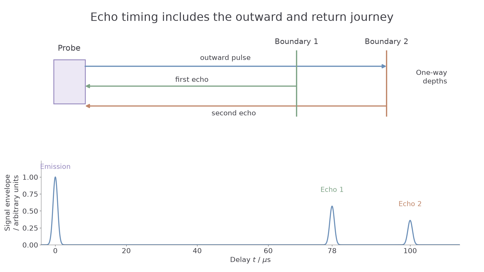
The delay is measured from emission to reception. For a boundary directly along the beam at depth , sound travels to the boundary and back. In one uniform medium, the distance is .
Pulse-echo distance
Here is one-way depth in , is sound speed in the medium in , and is the round-trip delay in . The pulse follows the same outward and return path, and the speed is treated as constant. For different layers, add the travel times in those layers instead of using an unjustified single speed.
Not on the data sheet: learn it.
[DIAGRAM: asset_name: Ultrasound paths and echo delays; asset_slug: pulse-echo-paths; caption: Each echo delay includes the outward and return sound paths; description: A probe sends a pulse along a straight beam towards two planar tissue boundaries. Outward and return arrows are separated for clarity but represent the same beam path. A companion signal trace shows emission at zero and echoes at 78 and 100 microseconds. Depths are measured one way from the probe.]
Worked example 1: two boundaries
Question. Two echoes return after and . Treat the sound speed throughout as . Find the depth of the nearer boundary and the separation of the boundaries.
Plan. Halve each relevant round-trip distance. The difference in echo times gives the extra outward-and-return travel between boundaries.
Answer. The first boundary is about deep, and the separation is about .
Check. The second depth is , so subtracting the two depths gives the same separation. Omitting the factor of two would double both answers.
Worked method: two different sound speeds
A pulse crosses a first layer of thickness at , then a second layer at , and returns from its far face after . First remove the time spent crossing the known layer twice:
Only this remaining time belongs to the second layer, so its thickness is . Using one sound speed for the whole journey would assign the wrong time to at least one layer. Adding the two round-trip times checks the result.
Scanning the beam across the body builds up boundary positions. Echo signals can be displayed as brightness at their inferred depths to form an image. A single line of echo delays is not already a complete cross-sectional picture.
Short pulses also help distinguish boundaries close together along the beam. If a pulse lasts , echoes with delays differing by much less than this overlap. In a simple pulse-length model, axial separation needs to be about or greater. A pulse in a medium with speed corresponds to about separation. This is a pulse-length estimate, not a complete model of scanner resolution.
Watch out
The frequency's period and the complete pulse duration are different. A burst containing several cycles lasts several periods. Shortening the whole burst reduces echo overlap.
Impedance mismatch controls reflection
Why does gel help sound enter the body? An air gap gives a very large mismatch between the acoustic properties on its two sides, so nearly all the incident intensity is reflected. Coupling gel removes that gap and provides a much closer match to tissue.
Specific acoustic impedance
The specific acoustic impedance of a medium is the product of its density and the speed of sound in that medium.
Continue this lesson.
You’re previewing NovaLearn without an account. Create a free account to read the rest.
Create free account