22.3 - Wave-particle duality and electron diffraction
Connect wave and particle evidence for light and matter, explain an electron-diffraction pattern, and calculate de Broglie wavelengths from momentum and accelerating potential difference.
Wave and particle evidence
An electron beam produces diffraction rings: how can something with mass and charge show the behaviour of a wave? The evidence tells us that a purely classical particle picture is incomplete, just as a purely classical wave picture was incomplete for photoelectric emission.
Light's interference and diffraction demonstrate its wave behaviour. Photoelectric emission demonstrates discrete photon energy exchange: an electron needs enough energy in a single absorbed photon, so frequency has a role that increased intensity alone cannot replace.
Electrons also show complementary aspects. They can be detected in individual localised interactions, carry definite charge and transfer momentum. Yet a beam can produce an interference or diffraction pattern whose positions depend on an associated wavelength.
Wave–particle duality
Wave–particle duality describes the evidence that electromagnetic radiation and matter exhibit both wave-like and particle-like behaviour.
This does not mean that an electron is a tiny classical ball that physically changes into a water wave whenever convenient. It is a quantum object. Different experiments reveal different aspects, and neither classical picture alone describes all the observations.
You already know
A diffraction pattern has an organised distribution of maxima and minima. It is the interference of wave contributions that explains those bright and dark regions, rather than simply a uniform spreading of particles.
For light, use observations of electromagnetic radiation as evidence. For matter, use observations involving particles such as electrons. Electron diffraction is evidence for the wave nature of matter, not another example of a light wave's diffraction.
You do not need a new formula to identify the evidence, but you do need to say which physical object and which behaviour the experiment tests.
Electron diffraction through a thin film
A typical demonstration accelerates electrons in an evacuated tube and directs them through a thin polycrystalline film, such as graphite. A fluorescent screen beyond the film shows bright rings around the central beam.
The crystal contains regularly spaced atoms. Wave contributions associated with scattering from that structure interfere, giving strong intensity in particular directions and weaker intensity between them. Many small crystallites with different orientations can produce concentric rings rather than one simple set of parallel fringes.
[DIAGRAM: asset_name: Electron diffraction in an evacuated tube; asset_slug: quantum-electron-diffraction; caption: An accelerated electron beam produces rings after passing through a thin crystalline film; description: A side-view evacuated tube contains an electron source, accelerating anode, thin polycrystalline film and fluorescent screen. The central beam and symmetrically diffracted directions reach the screen. A separate front view shows concentric bright rings and a central spot. Accelerating potential is labelled between source and anode, and the screen view is identified as an electron-intensity pattern rather than literal circular electron paths.]
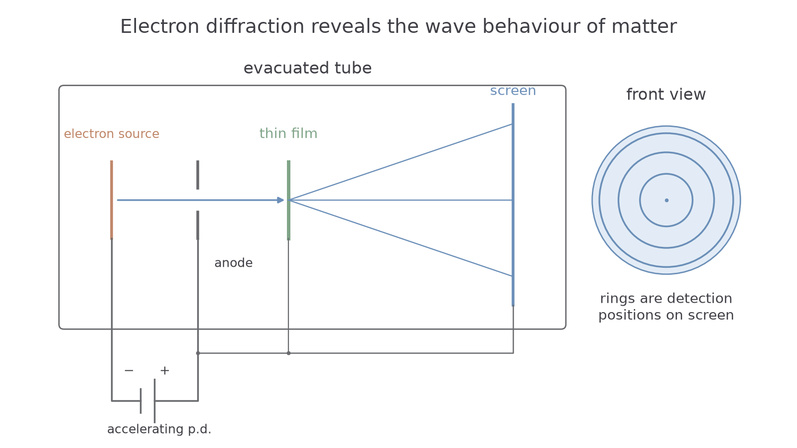
A classical straight-line trajectory picture alone does not account for the organised diffraction pattern and its wavelength dependence. The pattern is evidence that the moving electrons have wave behaviour. It is not evidence that the electron's charge has disappeared.
The bright rings mark where many electrons are detected. An individual arrival can be localised while a collection of arrivals builds a wave-governed intensity pattern. Do not draw the electrons themselves travelling around the rings: the rings are locations on the screen.
Increasing the accelerating p.d. increases electron momentum. The associated wavelength becomes shorter, so the corresponding diffraction angles, and ring radii for the same film and screen geometry, become smaller. The next part derives the wavelength change quantitatively; detailed crystal diffraction formulae are not needed for this qualitative observation.
Model limit
The wavelength is associated with electron motion; it is not a wavelength of electromagnetic radiation emitted by the electron gun. The pattern concerns the probability distribution of electron detection in this setup.
Describe the evidence as a connected experiment, rather than as the word “diffraction” on its own.
Continue this lesson.
You’re previewing NovaLearn without an account. Create a free account to read the rest.
Create free account