Core practical 17 - Exercise and spirometer traces
Exercise changes both the depth and frequency of breathing, but moving air is different from consuming oxygen. In Core practical 17, use calibrated spirometer traces to measure tidal volume, breathing rate, respiratory minute ventilation and oxygen consumption, then judge how safely collected data support a comparison before and after exercise.
What the spirometer measures
A spirometer records the movement of gas into and out of a person's lungs, allowing breathing to be measured quantitatively.
Tidal volume
The volume of gas breathed in or out in one normal breath, usually expressed in litres or cubic centimetres.
Breathing frequency, also called breathing rate, is the number of complete breaths per minute. Respiratory minute ventilation is the total volume moved into or out of the lungs per minute. Do not add inspired and expired volumes together: they represent opposite movements of approximately the same tidal gas. Oxygen consumption is the volume of oxygen taken up by the body per unit time, usually expressed in litres per minute.
In one suitable school apparatus, a person breathes through a mouthpiece and one-way valves connected to a freely moving, water-sealed gas chamber. A pen or movement sensor records chamber movement against time. Expiration adds gas to the chamber and inspiration removes it. A nose clip and a good mouthpiece seal prevent unmeasured breathing through the nose or leaks around the lips. A modern flow sensor can instead measure flow and integrate it to obtain volume; its trace convention must be checked.
The model traces in this lesson show gas volume in the spirometer, not absolute volume inside the lungs. On these axes an upward movement represents expiration and a downward movement inspiration. Some instruments reverse the displayed direction. Read the axis and calibration rather than memorising that every upward stroke means inspiration. Ordinary spirometry does not directly measure the gas remaining in the lungs after maximal expiration.
For oxygen-consumption analysis, the apparatus must be a suitable closed system with carbon dioxide removed from expired gas by an approved absorbent. Oxygen entering the blood is used in aerobic respiration; exhaled carbon dioxide is trapped rather than replacing it in the chamber. The chamber's mean gas volume therefore decreases. Breath oscillation and this slower decrease provide different measurements. A simple open flow spirometer without gas analysis cannot measure oxygen consumption from a downward volume trend.
Safe consented participation
Human participation needs informed consent, a suitable activity and direct teacher supervision throughout recording.
The teacher explains the purpose, exercise, mouthpiece procedure, expected sensations and right to stop before asking for voluntary consent under the centre's rules. A learner may choose supplied trace analysis instead, without pressure to take part physically. Use participant codes, keep health information private and avoid public comparisons of fitness. Breathing data are not a diagnosis.
The teacher checks suitability using the centre's risk assessment and relevant medical advice. A current respiratory infection, an acute breathing problem, a relevant heart or lung condition, recent surgery, previous exercise-related fainting or another medical restriction can make participation unsuitable. Students do not decide that another person's condition is safe. If suitability is uncertain, use supplied traces. Do not withhold prescribed medication or impose fasting for this classroom investigation.
Each person needs a new disposable mouthpiece or one cleaned and disinfected by the approved manufacturer/centre procedure. Clean reusable tubing, valves and nose clips as specified, and wash hands. Use compatible filters where the apparatus procedure requires them. Sharing an untreated mouthpiece can transfer microorganisms in saliva or respiratory material. Do not improvise a filter that adds unsafe breathing resistance.
Use an approved, maintained spirometer. The teacher or technician checks valves, leaks, gas supply and absorbent before use. Carbon-dioxide absorbents can be corrosive and dusty: trained staff handle them with appropriate eye protection and prevent dust entering the breathing circuit. Oxygen supports combustion; trained staff secure and operate cylinders, keep ignition sources away and keep oils and grease off oxygen fittings. Follow the apparatus-specific safe recording duration and gas limits. A depleted oxygen supply may become dangerous without a clear warning sensation when carbon dioxide is absorbed, so comfort alone is not a safety monitor.
Exercise is a modest, teacher-approved activity on clear flooring with suitable footwear and stable equipment. Exercise takes place off the breathing circuit; the participant then sits for the post-exercise trace. Stop exercise or recording immediately if requested, or if there is dizziness, chest pain, unusual breathlessness, distress, impaired alertness or resistance to breathing. The supervisor returns the participant to room air and follows the centre's response procedure. Never continue to obtain a longer graph. These safeguards also apply when the intended measurement is only a short recording. Practical Biology's apparatus guidance supports supervised school use; ATS/ERS guidance provides additional infection-control and contraindication background for clinical spirometry.
Calibration and a controlled method
A valid comparison needs known volume and time scales and the same measurement conditions before and after exercise.
Before connecting a participant, trained staff check that a known syringe volume produces the correct volume change, following the instrument's calibration procedure. For example, if adding moves a pen by , the volume scale is per millimetre. A excursion then represents . Check across the working range rather than assuming one point proves every reading accurate. Verify the logger clock or chart speed against known time. At a chart speed of , represents , not one minute.
For closed-system post-exercise recording, trained staff prepare the oxygen supply and effective carbon-dioxide absorbent according to the approved apparatus procedure, checking and replenishing them before later recordings as required. The model graph's starting gas volume is an analytical example, not a prescribed fill volume or safety limit. Monitor gas temperature and pressure and retain the same reporting conditions for paired traces.
An appropriate paired protocol proceeds as follows, within the approved safe recording limits:
- Record the participant code, activity, timing, apparatus settings, gas conditions and room conditions. Allow the seated participant to rest and become accustomed to breathing through the mouthpiece while initially connected to room air.
- Under the teacher's control, switch to the prepared circuit at the end of a normal outward breath and collect a short calibrated resting trace of quiet tidal breathing, for example a 30-second window if approved for this apparatus. Avoid speech, deliberate deep breaths and unnecessary movement. Keep the raw trace and note coughs or leaks.
- Disconnect from the circuit. Carry out the approved activity for a fixed duration at a fixed pace, for example a modest step activity timed with a metronome. Keep step height, duration and pace the same across matched trials.
- Seat the participant again and begin the post-exercise window after the same measured delay each time. A stated five-second delay is a design example, not a universal safe requirement. Recording length, posture, mouthpiece seal and instrument settings match the resting measurement.
- Allow full recovery before a fresh paired trial. Record at least three matched trials where safe and feasible, rather than calling three breaths within one trace three independent trials. Use further consenting participants if investigating how widely the result applies.
The independent variable in a rest-versus-exercise comparison is exercise condition. The four dependent measurements are tidal volume, frequency, minute ventilation and oxygen consumption. Keeping the same participant in each pair limits differences in body size and physiology. Keep the activity and its duration, time to recording, posture and environmental conditions consistent. Do not confuse controlling activity across repeats with removing exercise as the intended changed condition.
Software can collect time-stamped volume readings, display calibrated axes and help count cycles or fit a trend. Save raw data alongside processed results, label units, record excluded segments with a reason and check the program's chosen breath boundaries. Automated output cannot repair a leak or identify an inappropriate participant. Analysing supplied traces develops the same interpretation skills without exposing a participant to the procedure; practical competence and the separate endorsement still require centre observation and authentication.
Tidal volume and breathing frequency
The vertical excursion of a trace measures the size of a breath, while its repeated cycles measure breathing frequency.
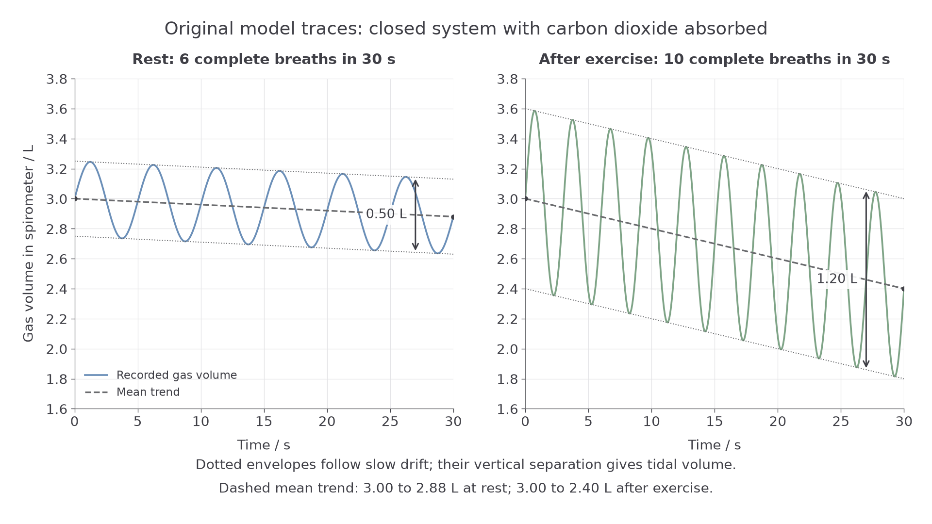
Original model traces on identical scales. The vertical axis is gas volume in the spirometer in litres; the horizontal axis is elapsed time in seconds. The dotted envelopes follow slow drift, while the dashed line shows the mean trend.
To measure tidal volume, find the volume difference corresponding to one normal breath, using the calibration. Where the whole trace drifts, compare the upper and lower envelopes at the same time, or remove the slow baseline drift before measuring excursion. A raw peak and a later trough have both a breathing difference and a small drift difference. Do not mistake the distance from the central trend to one envelope for the full tidal volume: it is half the excursion in these symmetrical model traces.
In the resting trace the envelopes are apart, so tidal volume is . After exercise they are apart, equivalent to . In real traces, measure several representative breaths in a stable window and calculate their mean instead of selecting the largest breath.
Here is breathing frequency in breaths per minute, is the number of complete breaths and is the recording interval in seconds.
Count complete cycles between equivalent phases. Six complete cycles contain six breaths, even though counting endpoints carelessly may produce seven boundary marks. The model trace contains six complete rest cycles in 30 seconds, giving breaths per minute. Ten complete post-exercise cycles give breaths per minute. Alternatively, divide 60 seconds by the mean period of one breath: the five-second resting period gives the same frequency. Wider excursions and more cycles show deeper and faster breathing respectively; neither is a reason to relabel the vertical axis as oxygen consumption.
Continue this lesson.
You’re previewing NovaLearn without an account. Create a free account to read the rest.
Create free account