2.1 - Gas exchange and Fick's law
Oxygen must cross an exchange surface before the blood can deliver it to respiring tissues. This lesson explains how surface area, diffusion distance and concentration difference determine that transfer, uses model-cell geometry and Fick's law to calculate their effects, and connects them to the structure of the mammalian lung.
What makes an exchange surface effective
An exchange surface separates two regions between which substances move. In gas exchange, oxygen and carbon dioxide cross it by diffusion.
Diffusion
The net movement of particles from a region of higher concentration to a region of lower concentration, due to their random motion.
Particles move in both directions, but more cross from the higher-concentration side, giving a net transfer. At equal concentrations there is no net transfer, although individual particles still move. Diffusion itself is passive: cells do not supply ATP to drive the particles across the surface. Breathing muscles can use energy to maintain the conditions for diffusion without turning gas diffusion into active transport.
Three properties explain efficient exchange.
| Property | Why it increases transfer per unit time |
|---|---|
| Large available surface area | More particles can cross at the same time. |
| Thin exchange barrier | Particles travel a shorter distance across it. |
| Large concentration difference across the barrier | A greater imbalance between the two directions of movement produces faster net transfer. |
The rate of diffusion is the amount transferred per unit time. The concentration gradient describes the concentration change over a distance. For the same concentration difference, a thinner barrier gives a steeper gradient.
Surface area must also be considered relative to demand. If each unit of living tissue has a similar oxygen requirement, a larger tissue volume requires more oxygen. A large surface area to volume ratio, written SA:V, provides more exchange area per unit volume. A small organism may exchange gases across its body surface; a large mammal needs specialised exchange surfaces connected to mass transport. Its blood carries oxygen from the lungs to internal tissues, so diffusion does not have to span the whole body.
Efficient exchange needs enough area for the tissue's demand, a short diffusion path and a maintained gradient. A large absolute area alone does not establish a large SA:V.
Model cells, surface area and units
Simple shapes let us compare how cell dimensions affect exchange, provided we state which surfaces are exposed. These are mathematical models, not claims that every real cell has a regular shape.
For a rectangular prism of length , width and height , the opposite faces form three equal pairs. Their areas are , and . Adding all six faces gives the external surface area.
For a rectangular prism with all six faces exposed:
Use the same length unit for , and .
Consider an idealised cell measuring .
SA:V can be written as , or , when the area is measured in square micrometres and volume in cubic micrometres. The quotient has units of inverse length; it is not a dimensionless number. Ratios calculated using different length units must be converted before comparison.
A flattened model of the same volume measuring has:
Its SA:V is , about times the first model's value. The shortest surface-to-centre distance also falls from to . Flattening can therefore increase exchange area per unit volume and shorten the path to the interior. This explains why a thin shape can be useful even when volume does not change.
Squared and cubed conversions
A length conversion factor must be squared for area and cubed for volume. Since :
Thus , whereas . Dividing by these factors reverses the conversions. Similarly, , so and . The prism's is ; its is .
Spheres, cylinders and the effect of size
Round and elongated model cells show that the same relationship between size and exchange applies beyond rectangular prisms.
For a sphere of radius , its surface area is and its volume is . Dividing cancels both and two powers of :
A spherical model cell of radius has and . Its SA:V is exactly for the stated model dimensions. At radius , area is , volume is , and SA:V is . The larger cell has more area, but only half as much area per unit volume.
For an idealised rod-shaped cell, use a closed cylinder of radius and length . Each circular end has area . Unwrapping the curved surface gives a rectangle with width equal to the circumference , and length . Its curved area is therefore .
For a closed cylinder with the curved surface and both ends exposed:
An open tube or a cylinder with inaccessible ends has a different available exchange area.
With and , circumference is ; each end has area . The curved area is , so total area is . Volume is , and SA:V is . Keep unrounded intermediate values, or cancel , when finding the ratio; round the final reported answer. The radius, not the diameter, belongs in these equations.
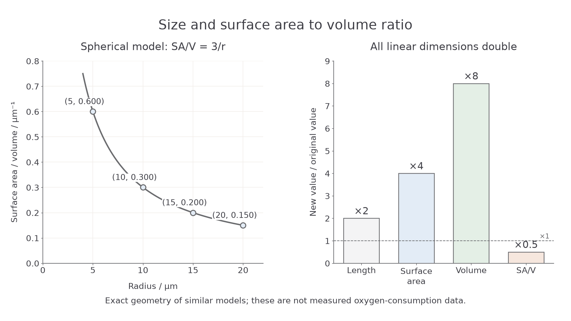
For spherical model cells, SA:V equals . The right panel shows the changes for any geometrically similar model when every linear dimension doubles.
At radii and , the graph gives and : doubling radius halves SA:V. Increasing radius from to halves it again to . This is an inverse curve, not a straight-line decrease.
When all linear dimensions double, area increases by , volume by , and SA:V becomes of its original value. The surface-to-centre diffusion distance also doubles. This conclusion applies to similar prisms and cylinders as well as spheres. Changing only one dimension, or flattening a cell, is a different comparison.
These exact geometric predictions explain an exchange constraint, but they do not prove that one real species consumes oxygen faster than another. Metabolic demand, temperature, permeability and the fraction of the surface actually available also matter.
Fick's law explains the rate
Fick's law brings the three exchange-surface properties into one relationship. Let be the available area, the diffusion distance across the barrier and the concentration difference between its two sides.
Under otherwise unchanged conditions:
The symbol means “is proportional to”. Doubling or doubles the rate; doubling halves the rate.
This is a relationship between quantities, not an unqualified equality. The proportionality assumes the same diffusing substance, medium and temperature. Different gases or barriers can have different diffusion properties. The area in Fick's law is the available exchange area, not the SA:V quotient. SA:V helps compare supply with tissue demand; it does not replace in the rate equation.
If concentration on one side is and on the other is , then . It is the difference, not either concentration alone, that matters. If both sides increase by the same amount, this difference stays unchanged. For a barrier thick, the model gradient has magnitude . Making it thick with the same halves the gradient and the rate.
For two model exchange surfaces with the same diffusion constant, let their rates be and :
Suppose available area falls to of its original value, the concentration difference stays the same and thickness doubles. The rate becomes of the original: remains, a decrease. Do not add a area loss to a thickness effect; their factors multiply.
Symbols keep comparisons precise. We write for a reduced rate, and for the same comparison reversed. The symbols and mean “much less than” and “much greater than”; for example, the short alveolar diffusion distance is the distance from a lung to a foot, while that body-scale distance is the alveolar distance. Equality gives an exact stated relationship; gives an approximation, such as . The specification also uses for “approximately”. A proportionality cannot yield an absolute rate unless a suitable constant is known.
Calculating and rearranging a diffusion model
A supplied diffusion constant allows Fick's proportionality to become an equation for an idealised barrier.
For a model with a constant diffusion coefficient :
Use in , in , in , in and in . The units cancel to moles per second. These examples use invented model values with concentrations expressed consistently for the model. They are not measured lung data or a way to calculate a patient's oxygen transfer from an alveolus sketch.
A rate calculation and estimate
Take , , and . Convert before substitution.
An estimate using gives:
Using the supplied values:
This is , since . It agrees with the estimate. Forgetting to convert micrometres would make the denominator one million times too large and the calculated rate one million times too small.
Finding the barrier thickness
Suppose the same , and give . Rearrange before substituting:
The rate has halved, so the calculated thickness should double. This independent proportional check makes the result more convincing than a calculator display alone.
The alveolar gas exchange surface
The mammalian lung places a large, delicate exchange surface inside the body while keeping it accessible to both air and blood.
Air passes through the trachea, bronchi and smaller bronchioles to the alveoli, tiny air spaces at the ends of the branching airways. Branching distributes air to many alveoli. Their numerous small air spaces provide a large total internal surface area within the lung volume; the alveolar surface, rather than the trachea, is the principal gas exchange surface.
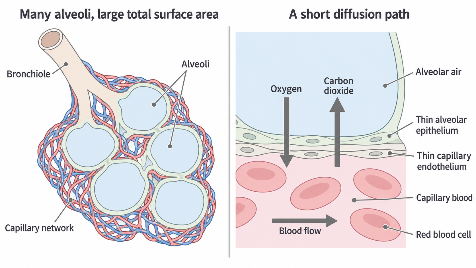
A conceptual view of the alveolar surface. Oxygen crosses from alveolar air to capillary blood; carbon dioxide crosses in the opposite direction. Both cross the thin alveolar epithelium and capillary endothelium. The layers are enlarged for clarity, not drawn to a measured scale.
An epithelium is a layer of cells lining a surface. Much of the alveolar epithelium consists of very thin, flattened cells, making it one cell layer thick with a short path through the cells. The nearby capillary wall is also one thin cell layer, called the endothelium. These two walls are closely apposed, with thin supporting material between them. Oxygen therefore travels only a short distance from air to blood, and carbon dioxide a short distance in reverse. Saying only “one cell thick” is incomplete: the thickness and close arrangement of both walls explain the short overall barrier.
A thin moist film lines the alveolar surface. Gases dissolve in this film before crossing the cells. Moisture enables transfer, but a thick accumulation of fluid would lengthen the diffusion path and reduce the rate. A dense capillary network places blood close to a large area of the alveolar surface.
The lung also has elastic tissue, allowing it to expand during inhalation and recoil during exhalation. Alveolar surfactant reduces surface tension in the lining film and helps prevent the air spaces from collapsing. Keeping alveoli open preserves available exchange area. Elastic recoil and surfactant support ventilation; neither directly pumps oxygen through the exchange barrier.
For an unfamiliar damaged surface, connect the changed structure to Fick's law. If walls between adjacent alveoli are lost, separate spaces merge and the total area available for diffusion falls. If the barrier thickens, increases. Either change reduces transfer rate when the other factors are unchanged, even if air still reaches the lung.
Keeping the diffusion gradients steep
A thin, large surface would still exchange gases poorly if air and blood were left unchanged until the gradients disappeared.
Ventilation and perfusion
Ventilation is the movement of air into and out of the lungs. Perfusion is blood flow through a tissue's capillaries; pulmonary perfusion carries blood past the alveoli.
Ventilation continually brings in air containing oxygen and removes air containing carbon dioxide. Inhalation involves contraction of the diaphragm and external intercostal muscles: the diaphragm flattens, the ribs move up and out, and thoracic volume increases. Lung expansion lowers air pressure inside relative to atmospheric pressure, so air flows in. During quiet exhalation these muscles relax; elastic recoil reduces lung volume, raises air pressure relative to the outside and moves air out. This is bulk movement of air driven by an air-pressure difference. Diffusion across the alveolar barrier is driven by a gas gradient.
Blood arriving through branches of the pulmonary artery has relatively little oxygen and more carbon dioxide after passing through respiring tissues. At the alveoli, oxygen diffuses into this blood and carbon dioxide diffuses into the air spaces. Continuous blood flow removes newly oxygenated blood and replaces it with blood that can receive more oxygen; it also delivers carbon dioxide for removal. Haemoglobin in red blood cells binds incoming oxygen, helping to keep the dissolved oxygen concentration low while uptake occurs. The blood then returns towards the heart through pulmonary veins.
Ventilation and perfusion therefore maintain the gradients from both sides. Ventilation replenishes alveolar oxygen and removes alveolar carbon dioxide. Perfusion carries newly absorbed oxygen away from the exchange region and brings further carbon dioxide in the arriving blood. If ventilation stops locally while blood continues to flow, alveolar oxygen diffuses into the blood without adequate replenishment and carbon dioxide accumulates; the gradients become less favourable. If blood flow stops while ventilation continues, fresh air alone cannot provide normal oxygen delivery to tissues because the transport route is missing.
The adaptations work together: many open alveoli provide area, closely apposed thin walls shorten the path, and moving air and blood maintain gradients. Increasing ventilation alone cannot fully compensate for a severely thickened barrier or loss of surface area; Fick's factors act together.