3.5 - Recognising organelles and measuring cells
An electron micrograph shows a thin slice through a real cell, so its structures look less tidy than a textbook diagram. In 3.5, you will learn to recognise the organelles named in 3.2 from their visible features, record observations carefully and calculate real sizes from calibrated images. You will also use simple geometry to estimate cell dimensions while keeping the assumptions clear.
Reading TEM evidence and recognising nuclei
A transmission electron microscope (TEM) passes electrons through a very thin specimen to reveal internal structures.
Resolution is the ability to distinguish nearby details as separate. An electron microscope can resolve much smaller structures than a conventional light microscope, so an enlarged TEM image can show membranes and ribosomes. Magnification alone cannot reveal detail that the instrument did not resolve. Dark regions in these prepared specimens usually indicate greater electron scattering, often associated with heavy-metal staining; they do not show the cell's natural colour.
A TEM section is a two-dimensional slice through a three-dimensional structure. A tube can look circular when cut across, elongated when cut lengthways and fragmented when cut obliquely. A section may miss a nucleolus or a centriole entirely. Absence from one slice does not establish absence from the whole cell. Preparation can also distort structures, so use several visible features and the specimen context rather than shape or darkness alone.
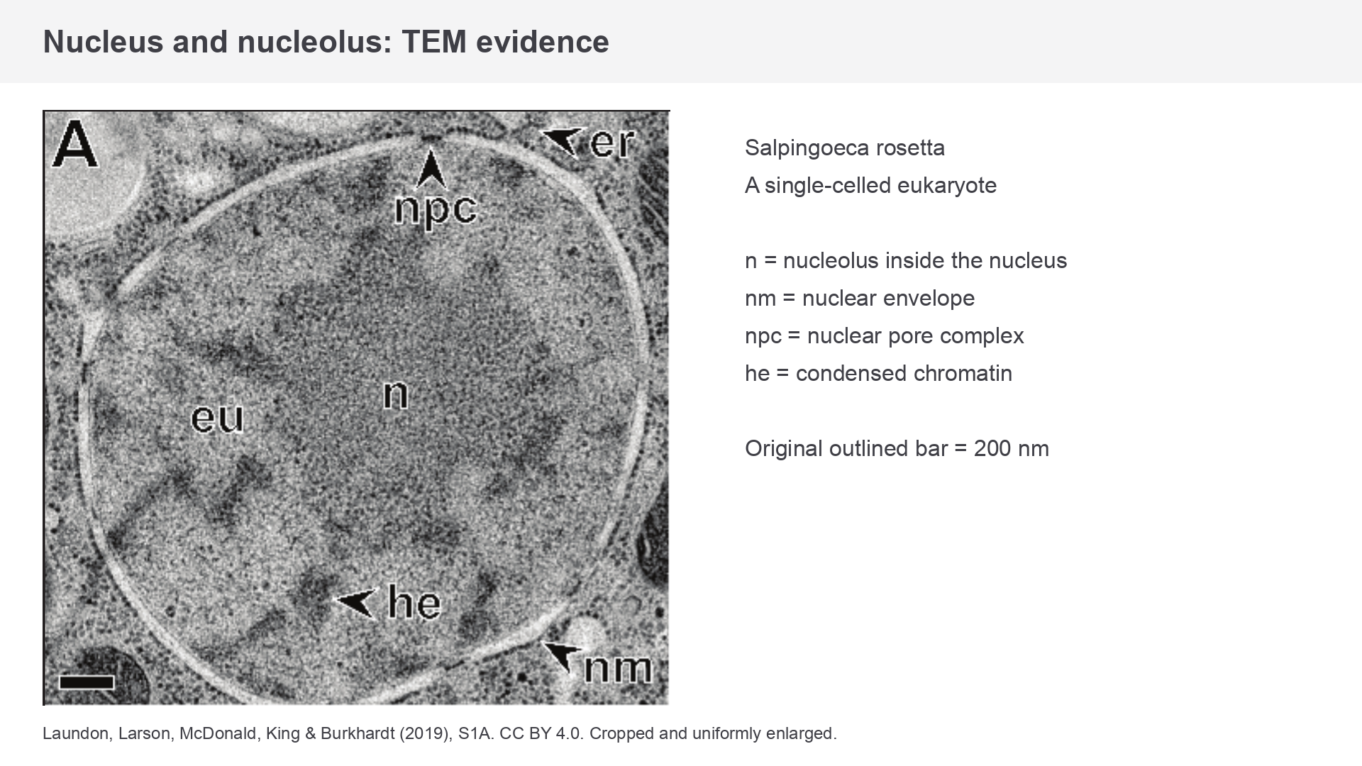
Figure 1. A nucleus in the single-celled eukaryote Salpingoeca rosetta. Source labels include n, nucleolus; nm, nuclear envelope; npc, nuclear pore complex; and he, condensed chromatin. Original outlined bar = 200 nm. Laundon, Larson, McDonald, King and Burkhardt (2019), S1A, CC BY 4.0; cropped and uniformly enlarged from the original research supplement.
The nucleus is a substantial region enclosed by a double nuclear envelope. Look for the boundary separating nucleoplasm from cytoplasm, variations in chromatin density inside it and, where the section and resolution allow, nuclear pores. The nucleolus is a denser region within the nucleus, without its own surrounding membrane. It is associated with rRNA production and ribosomal-subunit assembly. Do not label every dark nuclear patch as a nucleolus: condensed chromatin is also dark, so consider location, organisation and the source evidence.
Recording what you actually see
A scientific observation drawing records the specimen's visible arrangement, rather than a remembered ideal cell. On paper, choose a clear field, give a specimen/view title and use a large drawing with clear single outlines. Keep relative sizes and positions faithful to the field. Use a sharp pencil and straight, non-crossing label lines ending on the feature. Use annotations to add a relevant observation, such as “double boundary visible”; avoid decorative shading that hides boundaries. Include a calibrated scale bar, or a magnification valid for that exact drawing size. If no calibration is available, state that size cannot be determined; do not invent a bar.
For Figure 1, a drawing should distinguish the nuclear envelope from the nucleolus inside it and retain their relative positions. Do not add an unseen centriole, colour the nucleus as though natural colour were recorded, or turn every dark patch into a separate organelle. A drawing can communicate observed detail clearly, but cannot recover missing three-dimensional information.
Recognising ER and ribosomes
The endoplasmic reticulum is a membrane network, and its associated ribosomes help distinguish its rough and smooth regions.
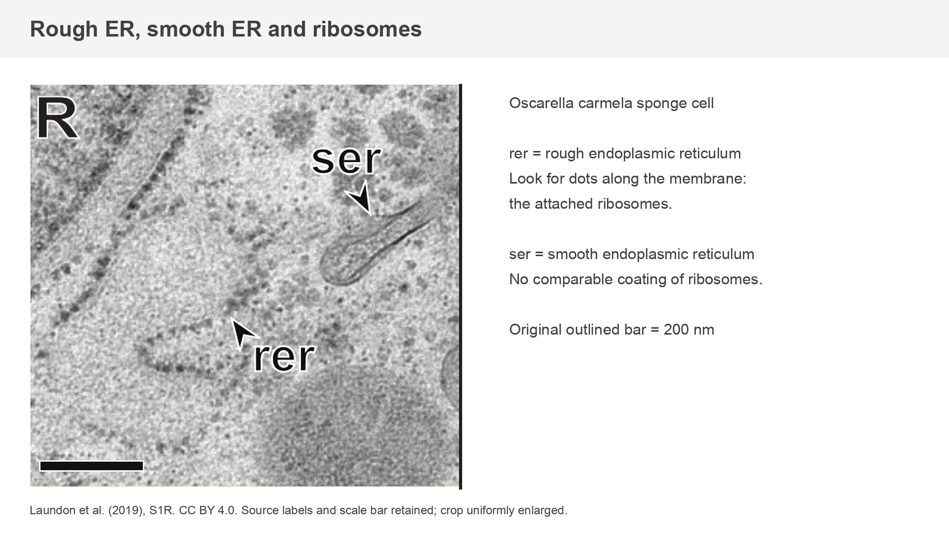
Figure 2. Oscarella carmela sponge cell: rer labels rough ER and ser labels smooth ER. The small dots on the rough ER membrane are ribosomes; the source arrows indicate the relevant profiles. Original outlined bar = 200 nm. Laundon et al. (2019), S1R, CC BY 4.0; cropped and uniformly enlarged from the research supplement.
Rough endoplasmic reticulum (rER) often appears as flattened membrane sacs, called cisternae, with small dense ribosome dots attached to their cytoplasmic surfaces. Trace the membrane around the paler interior, or lumen. Its “roughness” comes from attached ribosomes, rather than from the membrane being crumpled. Proteins made on these ribosomes can enter the rER for processing and transport.
Smooth endoplasmic reticulum (sER) lacks the attached ribosome coating. Its branching membrane tubules and sacs may appear as long profiles or small rounded sections. In Figure 2, follow the labelled ser profile and compare its surface with the dot-studded rer profile below. A smooth-looking isolated circle alone is insufficient: it could be a vesicle or a cross-section of a different tube. Continuity through further sections would strengthen identification.
Ribosomes are small, dense particles without a surrounding membrane. They occur free in cytoplasm, sometimes in groups, and attached to rER. Use their small size, repeated dot-like appearance and association with a membrane when identifying them. A much larger membrane-bound dark body is not a ribosome. Granules of stored material may also be dense, so “small dark dot” without size or context is weak evidence.
The useful comparison is therefore membrane arrangement plus ribosome distribution. It is stronger than “rER is dark and sER is pale”, because image contrast depends on preparation. These images teach recognition; the detailed protein-export pathway is developed in 3.3.
Recognising mitochondria and Golgi apparatus
A mitochondrion has internal membrane folds within one organelle, whereas the Golgi apparatus has a stack of separate flattened sacs.
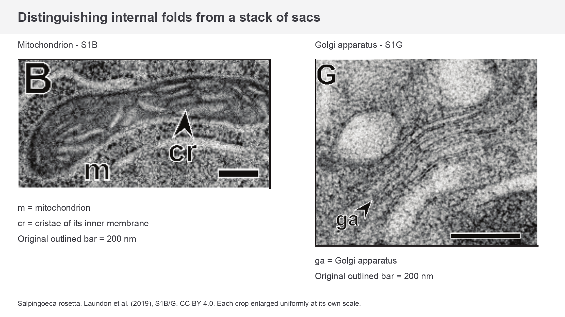
Figure 3. Salpingoeca rosetta: m labels a mitochondrion, cr its cristae, and ga the Golgi apparatus. Each original outlined bar represents 200 nm; the two crops have different enlargement factors, so compare each structure with its own bar. Laundon et al. (2019), S1B and S1G, CC BY 4.0; cropped and uniformly enlarged from the research supplement.
For a mitochondrion, look for an enclosing boundary and an inner membrane folded into cristae. The folds project into the matrix, increasing inner-membrane surface area for reactions involved in aerobic ATP production. A favourable high-resolution section shows both inner and outer membranes; a poorer image may not separate them everywhere. The entire organelle can look elongated, oval or nearly circular depending on shape and section direction. Cristae can also vary in appearance between species: the mitochondria in Figure 4 have a different internal profile from Figure 3.
The Golgi apparatus has a cluster of smooth, flattened, often curved cisternae, with small vesicles nearby. Unlike rER, its membranes do not carry a ribosome coating. Unlike mitochondrial cristae, the visible stack is not a series of folds inside a single double-membrane envelope. Nearby vesicles and the arrangement of several cisternae support identification; one curved line does not.
In an unfamiliar EM image, first trace boundaries, then inspect the internal arrangement and the surrounding structures. A reasoned identification might be: “mitochondrion, because internal cristae occur within its enclosing boundary”, or “Golgi apparatus, because several smooth flattened cisternae form a stack with associated vesicles”. A function such as “makes ATP” explains biological significance but is not itself a visible feature.
Recognising lysosomes and centrioles
Lysosomes are membrane-bound digestive compartments, while centrioles have an ordered microtubule structure.
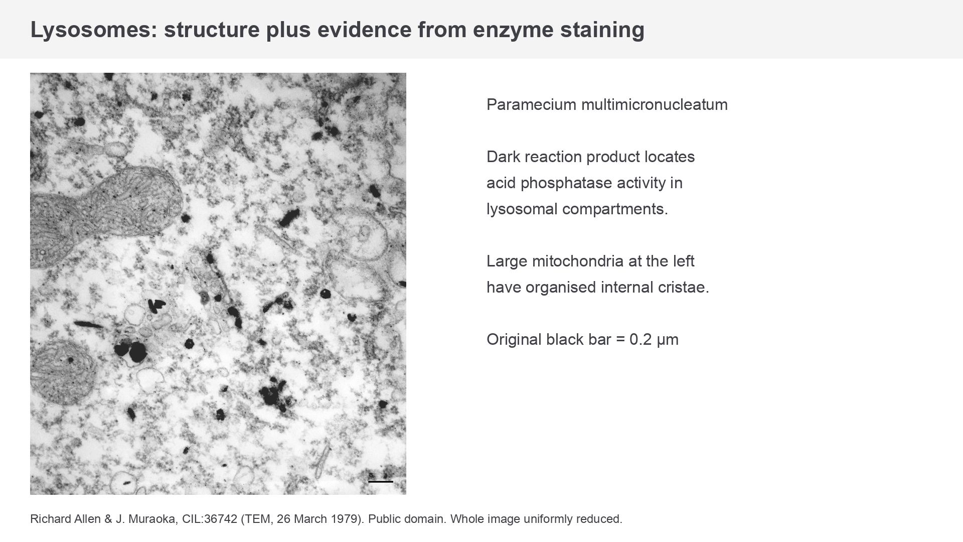
Figure 4. Dark reaction product provides evidence of acid phosphatase activity in lysosomal compartments of Paramecium multimicronucleatum. Large mitochondria with internal cristae are visible on the left. Original black bar = 0.2 µm. Richard Allen and J. Muraoka, TEM recorded 26 March 1979, Cell Image Library CIL:36742, public domain; whole image uniformly reduced. The repository describes this processed image as suitable for qualitative analysis.
A lysosome has one surrounding membrane and contains hydrolytic enzymes involved in intracellular digestion. In routine TEM it may appear as a rounded vesicle with dense or heterogeneous contents. Its appearance varies with the material being digested. A dark rounded profile could also be another vesicle or inclusion, so do not identify every dark circle as a lysosome.
Figure 4 includes additional evidence: a preparation locating the enzyme acid phosphatase. Dark reaction product in membrane-bound compartments supports their lysosomal identity. The enzyme evidence comes from the preparation and source description; darkness alone would not establish the enzyme's presence in an ordinary unstained or differently stained image. This illustrates the difference between an observation (“dense material inside a bounded profile”) and an interpretation (“a lysosomal compartment, supported by enzyme staining”).
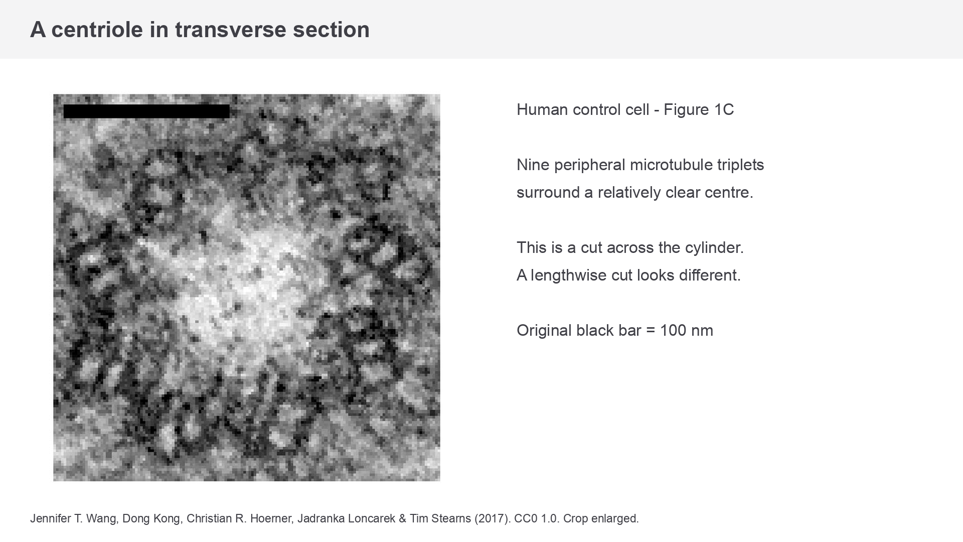
Figure 5. A human control-cell centriole in transverse section, cropped from Figure 1C of Wang, Kong, Hoerner, Loncarek and Stearns (2017). Original black bar = 100 nm. CC0 1.0; cropped and uniformly enlarged. The mutant-cell panels from the research figure are excluded.
A centriole is a short cylinder constructed from nine peripheral triplets of microtubules. A transverse section, as in Figure 5, shows the ring of triplets around a relatively clear centre. A longitudinal section shows a longer cylinder with roughly parallel microtubule profiles. Centrioles are often paired near the nucleus, but a thin section may show only one member or cut the two in different directions. They are not membrane-bound vesicles and do not contain cristae. Their characteristic microtubule arrangement provides stronger identification evidence than location alone.
Continue this lesson.
You’re previewing NovaLearn without an account. Create a free account to read the rest.
Create free account