2.7, 2.8 - Genes and the genetic code
The order of DNA bases carries information about the order of amino acids in a polypeptide. In 2.8, you will learn what a gene encodes; in 2.7, you will use the triplet, non-overlapping and degenerate genetic code to interpret that information. This explains how a four-letter base alphabet can specify many different amino acid sequences.
Genes and amino acid order
A protein-coding gene stores an ordered instruction in the base sequence of DNA.
Gene
A sequence of bases on a DNA molecule that codes for a sequence of amino acids in a polypeptide chain.
An amino acid is a building block of a polypeptide. A polypeptide is a chain of amino acids joined together. Different amino acids can occupy different positions along that chain: alanine–glycine–valine is a different sequence from valine–glycine–alanine, although both contain the same three types of amino acid. The detailed chemistry of joining and folding the chain is taught in 2.9.
DNA contains the bases adenine (A), thymine (T), cytosine (C) and guanine (G). Its information depends on their order, not just how many of each base it contains. Changing the order can change the instruction, just as rearranging letters can change a word. DNA bases are not amino acids and do not themselves become the polypeptide; the cell uses the information in their sequence to build the chain.
The genetic code is the set of rules connecting base sequences with amino acids. A gene is a particular stretch of DNA carrying information; the genetic code is the rule used to interpret that information. Two different genes can specify different amino acid sequences while using the same code.
This lesson concerns protein-coding genes, as in 2.8. Some other genes specify functional RNA rather than a polypeptide, so the definition should not be used to claim that all genes produce proteins. Nor does every stretch of DNA code for amino acids. NHGRI's gene glossary explains this broader use of the term.
Triplets and the reading frame
Three bases form each unit of the genetic code, allowing four base types to specify the 20 usual amino acids found in proteins.
The code is a triplet code: each group of three bases specifies one amino acid or a signal. On messenger RNA, these three-base units are called codons. Messenger RNA (mRNA) carries the coding information used when a polypeptide is made; it contains U (uracil) in place of DNA's T. The process that produces and reads mRNA is taught in 2.6. Here, the task is to interpret the sequence.
Why three bases? There are four possibilities at each position. A one-base code has only 4 different units, while a two-base code has 4 × 4 = 16: neither is enough to give a different code unit to each of the 20 usual amino acids. Three positions allow 4 × 4 × 4 = 64 different triplets. This gives enough possibilities for amino acid assignments and signals. It explains why a triplet code has sufficient capacity; it does not mean that a polypeptide contains 64 amino acids.
With four possible bases at each position, the number of possible sequences of length is . For a triplet, possible sequences.
The code is also non-overlapping. Once the starting position is fixed, bases 1–3 form one codon, bases 4–6 form the next, and bases 7–9 form the next. A base is used in only one of these successive codons. The second codon does not begin at base 2 and reuse bases from the first.
The reading frame is the way a sequence is divided into successive triplets from a specified starting position. For example, an internal mRNA section written 5′–GCUGAAGGU–3′, with the frame beginning at its first shown base, is read as GCU | GAA | GGU. The bars show boundaries; they are not extra bases or physical gaps. Starting from a different position would produce different groups, so a sequence problem must tell you the frame or supply enough information to establish it. The 5′ and 3′ labels distinguish the two ends of a nucleic acid strand; mRNA codons are read in the 5′ to 3′ direction.
Reading a codon table
A codon table lets you convert successive mRNA triplets into an ordered amino acid sequence.
This original teaching excerpt gives every standard-code mRNA codon beginning with G. The first and second bases select a row, and the third base selects a column. The entries name the amino acid; Ala means alanine, Asp aspartic acid, Glu glutamic acid, Gly glycine and Val valine. You need to use a supplied table accurately, rather than memorise these abbreviations.
| First two mRNA bases | Third base U | Third base C | Third base A | Third base G |
|---|---|---|---|---|
| GU | GUU → Val | GUC → Val | GUA → Val | GUG → Val |
| GC | GCU → Ala | GCC → Ala | GCA → Ala | GCG → Ala |
| GA | GAU → Asp | GAC → Asp | GAA → Glu | GAG → Glu |
| GG | GGU → Gly | GGC → Gly | GGA → Gly | GGG → Gly |
These assignments use the NCBI standard genetic code. The table is an excerpt, so an absent codon is outside this excerpt, not evidence that the codon has no meaning.
Worked sequence
The following is an internal coding section, with the reading frame beginning at the first shown base. It is not an entire gene or an entire mRNA, so it does not need to show the start and stop signals that lie outside the section.
The DNA coding strand is the strand whose sequence, written in the same 5′ to 3′ direction, matches the mRNA except that DNA has T where mRNA has U. It is a sequence label, not a statement that this strand is the template used to make RNA. Detailed strand roles are covered in 2.6. NHGRI's sequence-reading guide also distinguishes these strand labels and the reading direction.
| Sequence type | Internal section, written from 5′ to 3′ |
|---|---|
| DNA coding strand | 5′–GCT GAA GGT GTT GAC–3′ |
| Corresponding mRNA | 5′–GCU GAA GGU GUU GAC–3′ |
Read the mRNA left to right in its stated frame. GCU specifies Ala, GAA specifies Glu, GGU specifies Gly, GUU specifies Val, and GAC specifies Asp. The corresponding part of the polypeptide is therefore Ala–Glu–Gly–Val–Asp, in that order. There are 15 bases and five amino acids in this section; spaces just make the five non-overlapping triplets visible.
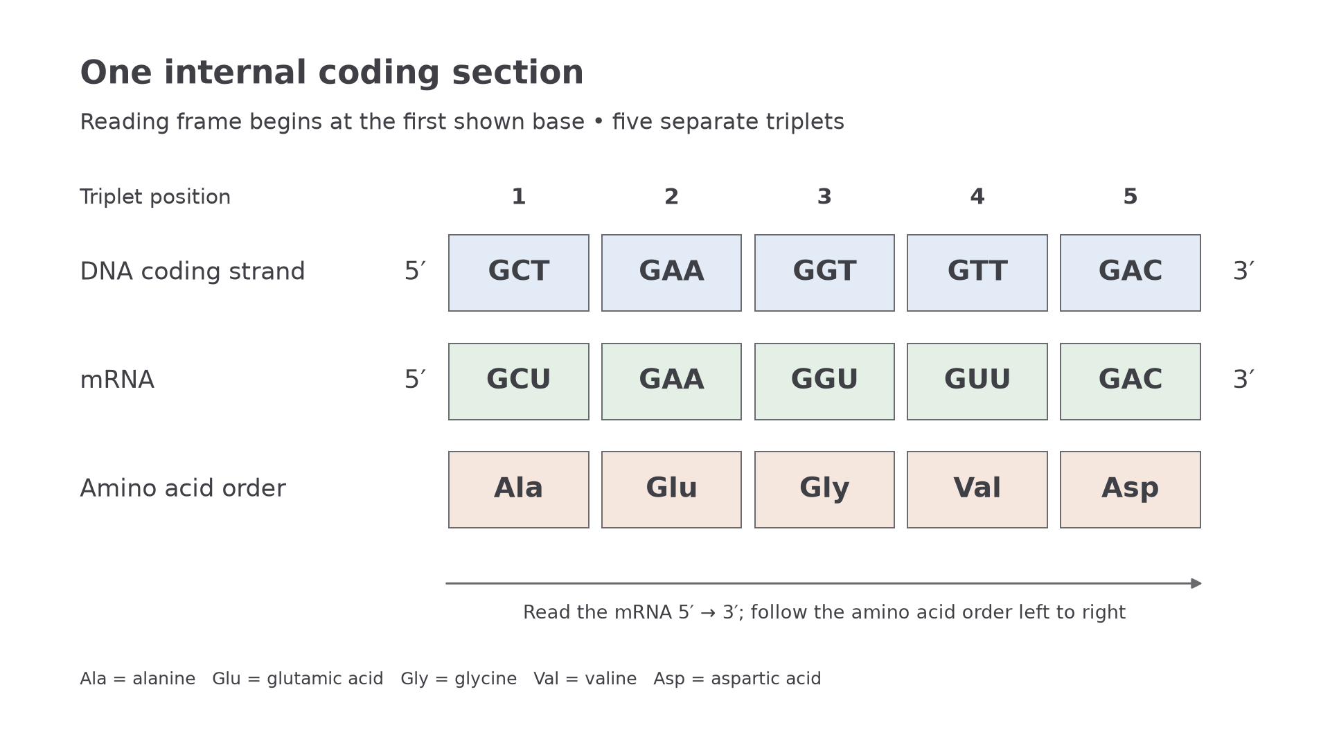
An original internal sequence example. Matching columns show how each labelled DNA coding-strand triplet corresponds to one mRNA codon and one amino acid position; the display follows the stated 5′ to 3′ direction.
The table above uses the RNA alphabet. For a supplied coding-strand DNA sequence written 5′ to 3′, replace T with U before using this mRNA table. Do not take its complement: that would answer a different strand question. For a supplied mRNA sequence, use its codons directly. Always check the sequence type, direction and frame before looking up the amino acids.
Degeneracy and sequence inference
Different codons can specify the same amino acid, which limits what you can infer from an amino acid sequence alone.
Degenerate genetic code
More than one codon can specify the same amino acid.
For example, GCU, GCC, GCA and GCG all specify alanine in the standard code. This is degeneracy: several different base triplets give the same amino acid assignment. It does not mean that one codon chooses randomly among several amino acids. When read in the stated frame under the standard code, GCU specifies alanine, not a choice between alanine and glycine. The assignment is unambiguous even though it is degenerate.
A consequence is that you can deduce an amino acid sequence from a correctly identified coding sequence and its code table, but you cannot usually reconstruct one unique DNA sequence from the amino acid sequence. A single alanine position could correspond to coding-strand DNA triplets GCT, GCC, GCA or GCG, each written 5′ to 3′. Two DNA coding sections can therefore have different bases yet specify the same amino acid order.
Do not turn this into the claim that every change at the third base leaves the amino acid unchanged. In the table, GAU and GAC specify aspartic acid, whereas GAA and GAG specify glutamic acid. Comparing GAU with GAA changes only the third base but changes the assignment. Check the actual triplets rather than assuming that a particular position never matters.
There are 64 possible triplets in the standard code: 61 specify amino acids, and three act as stop signals rather than specifying amino acids. Their detailed role, and the role of a start codon, belong to 2.6. Use the standard code for the examples here; code variants exist in some biological settings, so avoid claiming that the exact assignments are absolutely identical everywhere. NCBI records the standard and variant codes.
Triplet tells you the unit size; non-overlapping tells you how successive units use the bases; degenerate tells you that different units can have the same amino acid assignment.
These are three separate properties. None says that amino acid order is irrelevant, or that a codon table alone tells you the final shape and function of the protein.