1.5 - Atherosclerosis and blood pressure
Atherosclerosis develops in the wall of an artery before it can obstruct the blood flowing through it. In this lesson, you will follow how altered endothelial function, inflammation and plaque growth change an artery, then explain their effects on blood flow and pressure.
Endothelial dysfunction
The endothelium is the thin layer of cells that separates blood in an artery's lumen from the tissues of its wall.
The lumen is the space through which blood flows. The endothelium lines this space, with connective tissue and smooth muscle deeper in the arterial wall. A healthy endothelium is an active boundary: it controls movement into the wall, normally limits the attachment of circulating white blood cells, and releases signals that help smooth muscle relax. Relaxation allows a vessel to widen when appropriate; the endothelium is more than a smooth covering.
Endothelial dysfunction
A loss or alteration of the endothelium's normal protective functions, including its control of permeability, inflammatory cell attachment and vessel relaxation.
An affected region can become more permeable to lipid-containing particles in the blood. These particles carry fats, including cholesterol, and some can enter and be retained in the intima, the innermost region of the artery wall beneath the endothelium. The cells can also display more adhesion molecules: surface proteins to which particular white blood cells attach. Together, these changes make lipid retention and an inflammatory response more likely. Dysfunction does not require the whole endothelial layer to be scraped away or a visible hole to appear.
The protective role of this lining has experimental support. In isolated rabbit arteries, a chemical stimulus caused smooth muscle to relax when the endothelium was present. Removing the endothelium abolished that relaxation, showing that the lining participates in signalling to muscle rather than acting only as a passive surface. This experiment establishes a function of endothelium; it does not, by itself, demonstrate every step in human atherosclerosis. Furchgott and Zawadzki's arterial experiment.
Inflammation and foam cells
Lipid accumulation and altered endothelial signalling recruit inflammatory cells into the inner artery wall.
Inflammation is a response involving cell signals and the recruitment and activity of immune cells in affected tissue. In atherosclerosis, the response can persist without an infection. It is not simply a piece of fat sticking to the inside of a tube.
One recruited white-blood-cell type is the monocyte, which normally circulates in blood. Monocytes attach to the activated endothelial surface, pass between endothelial cells, and enter the intima. There they can develop into macrophages, cells that engulf material. Macrophages take up retained, modified lipid-containing particles. When large amounts of lipid collect as droplets in their cytoplasm, these cells have a foamy appearance and are called foam cells. A collection of lipid-rich cells forms an early fatty streak in the intima.
This sequence links three scales: changes in endothelial surface proteins affect the movement of whole cells; lipid uptake changes those cells; their accumulation changes the arterial tissue. Microscopy of arteries in a pig model identified monocytes at the endothelial surface, in endothelial junctions and within the intima, as well as lipid-containing phagocytic cells in lesions. These findings support recruitment from blood and development of macrophage foam cells in that model. Gerrity's original study.
Macrophages and other affected cells release chemical signals that can recruit and activate further cells. Continued lipid retention and inflammatory activity can therefore reinforce one another. Some foam cells eventually die, leaving lipid and cell debris in the wall. This helps explain why sustained inflammation can enlarge a lesion rather than clear it. It does not mean that every fatty streak progresses to an advanced plaque.
Plaque growth in the artery wall
A progressing fatty lesion becomes more complex as lipid, cells and fibrous tissue accumulate beneath the endothelium.
Smooth muscle cells can move from deeper in the arterial wall into the intima, multiply and produce extracellular material, including collagen, a strong fibrous protein. Collagen-rich tissue forms a fibrous cap over a deeper lipid-rich region that may contain dead cells and debris. The cap and core are different parts of the same lesion, not layers of a blood clot. Studies of human plaques have identified smooth muscle cells prominently in fibrous caps and macrophages in lipid-rich regions; studies of human arterial tissue also locate collagen production in lesions. Human plaque cell study, human collagen study.
Atherosclerosis
A disease in which lipid-rich plaques containing cells and fibrous tissue develop in the inner walls of arteries.
An atherosclerotic plaque, also called an atheroma, can thicken the intima and bulge towards the lumen. The remaining space for blood then becomes smaller. The wall can also become less flexible as its structure changes. These are two distinct effects: lumen narrowing alters resistance to flow; reduced flexibility alters how the artery responds to a pressure pulse. Early growth may be partly accommodated by outward enlargement of the artery, so plaque formation does not always cause immediate severe narrowing. Human coronary-artery enlargement study.
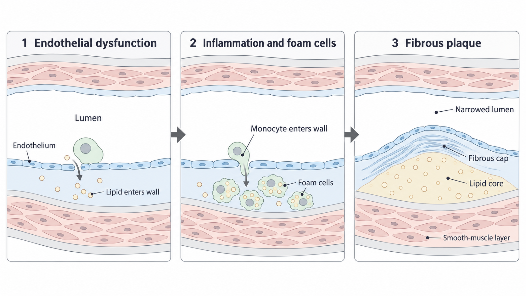
The plaque grows within the inner artery wall. Its fibrous cap lies beneath the endothelial lining and over the lipid core; the deeper smooth-muscle layer remains outside it. Arrows show a possible progression, not a fixed timetable or an inevitable outcome. This is a conceptual illustration, not a micrograph or a measured cross-section.
Notice that the endothelium follows the raised surface of the plaque. The plaque is not a loose lump travelling along the lumen. In a coronary artery, sufficiently severe narrowing can restrict the blood supply, and therefore oxygen delivery, to heart muscle, especially when its demand rises. The effect depends on the degree and location of the obstruction.
Some plaques have caps that weaken and rupture, exposing underlying material to blood. Other plaques remain unruptured. Rupture can trigger clot formation, which is a separate process from gradual plaque growth; the clotting mechanism is taught in lesson 1.6. NHLBI atherosclerosis overview.
Flow, resistance and blood pressure
Blood flow depends on the pressure difference driving it and the resistance offered by the vessel.
Blood pressure is the force per unit area exerted by blood on a vessel wall. Flow rate is the volume passing a point per unit time; it is not the same quantity as the speed of an individual blood cell. Resistance describes opposition to flow. A smaller lumen generally offers greater resistance, if other relevant conditions are unchanged.
The pressure difference is between the two ends of the vessel or region being considered. This is a simplified relationship for reasoning about flow; a real circulation is a branching, pulsating system.
The relationship gives two useful predictions. At the same pressure difference, increased resistance through a narrowed artery reduces flow. To maintain the same flow through that increased resistance, a greater pressure difference is required. Both conclusions follow from the relationship; neither means that pressure rises at every point inside the narrowed region. Pressure is lost along a resistive pathway, and the supply beyond an obstruction may fall. Cardiovascular haemodynamics.
For example, consider a vessel model connected to a pump. If plaque-like narrowing is added while the pump keeps the same pressure difference between the inlet and outlet, less fluid passes through per unit time. If the pump is adjusted to restore the original flow, it must generate a larger pressure difference. The stated condition determines the prediction; simply saying “the blood is squeezed, so pressure rises” misses that condition.
The large arteries normally stretch when they receive blood from the heart. A stiffer wall stretches less for a given increase in volume, so the pressure pulse can be larger. Systolic pressure is the highest arterial pressure during the heart's pumping cycle. Reduced arterial flexibility can contribute to raised systolic pressure. A longitudinal community study found that higher measures of aortic stiffness were associated with later blood-pressure progression. That finding is compatible with the mechanism, but the study did not establish that atherosclerotic plaques caused every participant's pressure change. Kaess and colleagues' follow-up study.
Raised pressure and atherosclerosis can also reinforce one another: sustained high pressure can place extra mechanical stress on the endothelium, promoting dysfunction, while vessel changes can contribute to higher pressure. A single local plaque does not guarantee high blood pressure throughout the body. Whole-circulation pressure also depends on the heart's output and the resistance of other vessels. NHLBI's explanation of arterial damage.
Plaque can restrict flow through a smaller lumen and can alter arterial flexibility. State what is held constant when connecting narrowing, flow and pressure; do not assume every plaque produces the same pressure change.
These distinctions allow you to predict a change without confusing a local obstruction with the pressure of the whole circulation.
Interpreting evidence for the mechanism
An observation supports a mechanism only when the feature measured is connected to the step being proposed.
For cell recruitment, finding monocytes attached to the luminal surface, passing through endothelial junctions and present in the intima supports a route from blood into the wall. Identifying lipid droplets in intimal macrophages supports lipid uptake and foam-cell formation. It is stronger reasoning to connect each finding to its proposed step than to say that a study “proves atherosclerosis”.
When reading a scientific report, first separate observations from interpretations. An observation is what the method recorded, such as a stained cell's location. An interpretation proposes what that observation means, such as the direction in which the cell moved. Define unfamiliar words from the supplied context: for example, a phagocytic cell can engulf material. Then connect the reported result to biology you have learned, while keeping the conclusion within the study's scope.
Worked interpretation
Gerrity's study used microscopy and cell-identification methods on arteries from a pig model. It reported monocytes on the endothelium, in its junctions and within the intima, together with phagocytic cells containing lipid droplets in lesions. The location and identity of these cells support monocyte recruitment and development into macrophage foam cells. This is a reasoned interpretation of the observations, not a direct recording of every cell's journey. Original monocyte study.
Fixed tissue samples capture particular moments. They cannot show the future course of each individual lesion, and findings from an animal model require care when generalised to humans. Several methods and studies can strengthen a mechanism by supporting different steps; no single image establishes the complete sequence, the timing, or the inevitability of rupture.
For pressure evidence, distinguish an association from a controlled comparison. People with stiffer arteries may later have higher pressure, but other differences between them could also affect pressure. By contrast, comparing flow before and after narrowing a model vessel while keeping its pressure difference unchanged isolates the effect of resistance more directly. The model supports the physical relationship, but it does not reproduce the full biology of a living artery. Match both the conclusion and its limitation to the actual method.