2.2 - Membranes and the fluid mosaic model
A cell membrane keeps the cell's contents separate from its surroundings while allowing selected substances and signals to cross. In 2.2(i), you will connect the arrangement of lipids and proteins to these properties. In 2.2(ii), you will see how measurements and experiments led scientists to develop, test and revise the fluid mosaic model.
Why phospholipids form a bilayer
The cell-surface membrane, or plasma membrane, surrounds the cytoplasm. Membranes also surround many cell organelles, keeping different reaction conditions in separate compartments. A membrane is a thin boundary made mainly of lipids and proteins, rather than a rigid wall.
A typical phospholipid contains a glycerol backbone attached to two fatty acids by ester bonds and to a phosphate-containing head. Compare this with a triglyceride, which has three fatty acids attached to glycerol. The phosphate-containing head is polar and hydrophilic, so it interacts with water. The fatty-acid tails are largely nonpolar hydrocarbons and hydrophobic, so they do not interact favourably with water.
Amphipathic
Having both a hydrophilic region and a hydrophobic region within the same molecule.
Water is present both outside the cell and in the cytoplasm. Phospholipids therefore arrange into a bilayer, two molecular layers: the outer layer's heads face the extracellular fluid, the inner layer's heads face the cytoplasm, and the tails of both layers point into the membrane's centre. This reduces contact between the hydrophobic tails and water. The heads do not point towards one another in the centre.
The phospholipids are separate molecules held in this arrangement by noncovalent interactions: attractions that do not join them into one giant molecule through covalent bonds. A small break exposes hydrophobic tails to water, so nearby lipids tend to rearrange and close the break. A bilayer can bend while maintaining a continuous boundary.
The hydrophobic interior is a barrier to most ions and large polar molecules. Small nonpolar molecules, including oxygen and carbon dioxide, can dissolve in the lipid region and cross more readily. Water can cross a lipid bilayer to some extent, and many membranes also contain water-channel proteins. Thus selective permeability means that different substances cross at different rates; it does not mean that only one substance can cross.
Fluidity and cholesterol
The bilayer has an organised orientation, but its molecules are not fixed in a lattice. Phospholipids move sideways within a layer, rotate and flex their tails. This lateral movement helps the membrane bend and allows its components to change neighbours. Movement from one leaflet to the other is much less frequent without assistance because a polar head would have to cross the hydrophobic interior.
Fluid mosaic model
A model in which a fluid lipid bilayer contains a varied arrangement of membrane proteins, some embedded in or spanning the bilayer and others associated with its surfaces.
“Fluid” describes movement within the membrane. “Mosaic” describes the mixture of different components. It does not mean that the proteins form a continuous sheet or that every component moves equally freely. A membrane can be fluid while remaining an effective boundary.
Temperature and lipid composition affect fluidity. Warming generally increases molecular motion within the range in which the membrane remains intact. Saturated fatty-acid tails can pack relatively closely. A cis double bond introduces a bend in an unsaturated tail, making close packing harder; a higher proportion of these tails tends to maintain fluidity at lower temperatures. These comparisons assume that other relevant conditions are similar.
Animal cell membranes also contain cholesterol. Its small polar hydroxyl group lies near the phospholipid heads, while its rigid hydrocarbon rings and nonpolar tail lie among the fatty-acid tails. Cholesterol therefore fits within a leaflet rather than forming a second layer outside the membrane.
Cholesterol modifies lipid packing. In a fluid membrane it restrains some tail movement and reduces permeability to many small water-soluble substances. At lower temperatures it also disrupts the close packing that would otherwise make the lipids more ordered and less fluid. It helps buffer changes in membrane properties; saying that it always increases, or always decreases, fluidity misses the dependence on conditions. Its effect on packing helps maintain a stable, selectively permeable boundary.
Fluidity concerns the movement and packing of membrane components. Permeability concerns how readily a substance crosses. These properties are related, but they are not interchangeable.
Membrane proteins and surface recognition
Proteins give membranes functions that a lipid barrier alone cannot provide. A protein consists of one or more polypeptide chains, whose amino acids are joined by peptide bonds. Different amino-acid side groups interact differently with water. The chain folds into a specific three-dimensional shape; the locations of its hydrophobic and hydrophilic regions help determine how it sits in a membrane.
An integral protein is embedded in the bilayer. A transmembrane protein spans it completely. Hydrophobic regions on the outside of its embedded portion contact the lipid tails, while hydrophilic regions project into the aqueous fluid on either side. A peripheral protein is associated with a membrane surface, often through interactions with other proteins, rather than extending through the hydrophobic core.
Two important transmembrane structures are channels and carriers. A channel protein has a water-filled pore, lined by hydrophilic groups, through which suitable ions or polar molecules can pass. The pore's size and chemical properties make it selective. A carrier protein binds a particular substance and changes shape, making its binding site accessible from the other side. It does not provide a continuously open pore. These structures explain why some substances can cross despite the hydrophobic barrier. The different transport processes and their energy requirements are developed in 2.4.
Other proteins act as receptors: a signalling molecule binds to a suitable site and triggers a response inside the cell. Some membrane proteins are enzymes, while others connect the membrane to internal support structures or help cells attach to one another. Their shape and location allow these functions to occur at a particular boundary.
A glycoprotein is a protein with an attached carbohydrate chain. A glycolipid is a lipid with an attached carbohydrate chain. In the cell-surface membrane, these chains project from the extracellular face. They contribute to cell recognition, adhesion and interactions with signalling molecules. For example, different carbohydrate structures at a cell surface can act as recognition markers. They do not form a second bilayer, and recognition does not require the whole glycoprotein to leave the membrane.
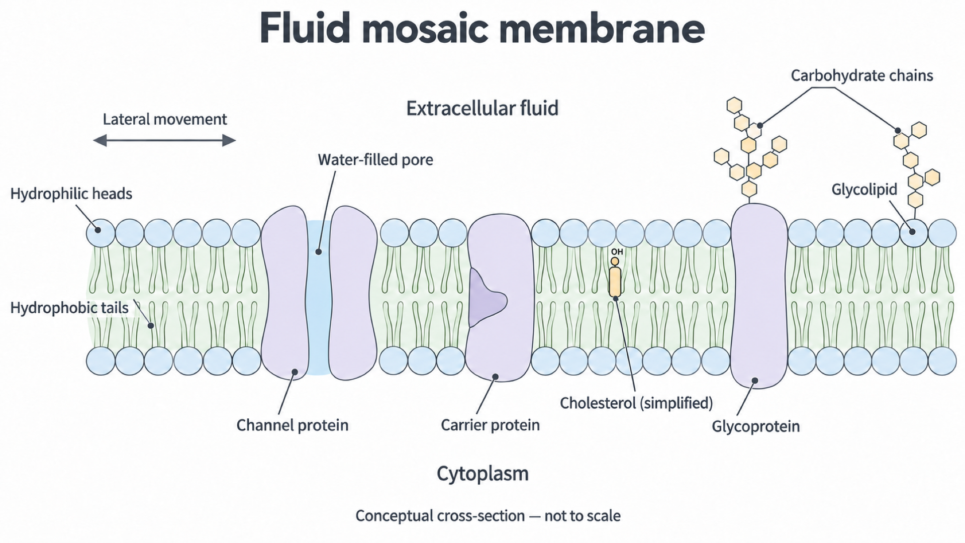
Conceptual cross-section of an animal cell membrane. The channel has a continuous water-filled pore; the carrier has no continuously open through-pore. Carbohydrate chains face the extracellular fluid. The cholesterol symbol shows orientation only, rather than its chemical structure; components are not drawn to scale.
Consider what happens if a channel's pore becomes blocked. The lipid bilayer can remain intact, yet passage of the channel's particular ion is reduced. By contrast, changing a receptor's binding site can reduce a response to a signal without necessarily changing the movement of that ion. Different membrane proteins give different functional consequences.
Lipid evidence for two layers
A model is an interpretation that explains observations and can be used to make predictions. Seeing a bilayer in a teaching diagram is therefore different from measuring evidence that supports two molecular layers.
In 1925, Gorter and Grendel extracted lipids from red blood cells and spread them as a single molecular layer on water. They compared this film's area with the estimated total surface area of the cells. The film area was approximately twice the cell surface area. If the lipids originally formed two layers over that surface, spreading them into one layer would give approximately twice the area. Their original report therefore supported a lipid bilayer.
Mature mammalian red blood cells were useful because they lack internal membrane-bound organelles. Extracted lipid was consequently less likely to include large amounts from internal membranes. The experiment measured lipid film area and estimated cell surface area; it did not directly photograph two rows of molecules or reveal the position of proteins.
For an original illustrative example, suppose the cells have an estimated total surface area of and their extracted lipid forms a monolayer of . The area ratio is
Both areas use the same units, which cancel. A ratio near two is consistent with two lipid layers, allowing for measurement uncertainty and assumptions about molecular packing. These are practice values, not a transcription of the historical results.
This inference depends on assumptions. Lipid should be extracted fully, the film should really be one molecule thick, and the cell surface area should be estimated accurately. Losing lipid during extraction would reduce the measured film area and lower the ratio. Underestimating cell surface area would also distort the ratio, but in the opposite direction: the denominator would be too small, so the ratio would be too high. A result near two is supporting evidence, rather than conclusive proof of every aspect of membrane structure.
Why protein coatings were revised
Lipid-layer evidence did not explain where membrane proteins were located. In 1935, Davson and Danielli proposed a lipid bilayer coated with protein on both surfaces, often called a sandwich model. This provided one explanation of membrane properties. Later thin-section electron microscopy showed two dark lines separated by a paler region, an appearance compatible with such a model.
Compatibility is not the same as a unique explanation. A dark line records how a prepared specimen affects the electron image; it is not a direct label saying “continuous protein sheet”. Different molecular arrangements may produce a similar pattern, and fixation or staining can affect the appearance. Evidence that fits two models cannot, by itself, distinguish them.
Freeze-fracture electron microscopy gave different evidence. A rapidly frozen membrane is fractured, often splitting through the bilayer's hydrophobic interior. A metal replica of the exposed fracture face is examined. Many such faces contain raised particles distributed within smoother regions. This observation is difficult to explain if all proteins form continuous coatings outside the bilayer, but fits a model in which proteins are embedded within it. Singer and Nicolson discussed this reasoning in their 1972 model paper.
The particles' appearance alone did not identify their chemical composition. Experiments combining independently identified membrane proteins with lipids strengthened the interpretation. In 1976, Yu and Branton combined the red-cell membrane protein Band 3 with lipid; the resulting artificial bilayer vesicles showed freeze-fracture particles resembling those in natural membranes. This was additional experimental support for the protein interpretation, obtained after the 1972 model had been proposed. It was not evidence available to its original authors in 1972.
The revised arrangement explains both membrane images and function: embedded proteins can form selective pathways across a lipid barrier, with hydrophobic regions contacting tails and hydrophilic regions contacting water. No single freeze-fracture image shows every protein's identity, function or movement. Structure and mobility require different kinds of evidence.
Evidence that membrane proteins move
A still image cannot establish whether a protein changes position over time. Frye and Edidin addressed mobility in 1970 by fusing cultured mouse and human cells, producing cells whose surfaces carried both species' antigens. They used fluorescent antibodies to distinguish the two kinds of surface marker.
At an early observation, around five minutes after fusion, the two markers were largely in separate regions. At , more than 90% of fused cells showed total intermixing of the markers within 40 minutes. Treatments inhibiting protein synthesis did not prevent this mixing, whereas lowering temperature inhibited it. These observations are reported in the original study's abstract.
The changing marker distribution supports movement of surface components. Continued mixing when protein synthesis was inhibited makes rapid production and insertion of new proteins a less satisfactory explanation. Lateral movement through a fluid membrane is consistent with the results and with the fluid mosaic model. The observation is the redistribution of fluorescence; lateral diffusion is the biological interpretation.
The authors considered alternative mechanisms, including movement of existing antigens into the cytoplasm and back to new surface positions. The mixing observation alone does not uniquely rule out that route. Nor does it show that every protein in every cell is freely mobile: the experiment followed particular antigens in particular fused cells. Stronger explanations come from agreement between different methods, not from calling one observation absolute proof.
Singer and Nicolson's 1972 model brought together lipid organisation, embedded proteins and mobility. A fluid bilayer with a varied protein arrangement accounted for more of the available evidence than a simple picture of fixed external protein coatings. The dates matter: the 1970 cell-fusion result could contribute to the 1972 model; the 1976 reconstitution result strengthened it later.
A useful model can still be revised
The fluid mosaic model remains useful, but an empty sea of lipids with every protein drifting without restriction is an oversimplification. Some membrane proteins connect to the cytoskeleton, a network of protein fibres inside the cell. Others interact with neighbouring proteins, external structures or particular lipids. These interactions can confine movement and create regions with different compositions.
More detailed tracking allows scientists to test this refinement. Fujiwara and colleagues followed individual phospholipids in rat kidney cells in a 2002 study. Many trajectories showed temporary confinement followed by movement to a neighbouring region, rather than unrestricted movement across the whole surface. Changes to the membrane-associated actin network altered this behaviour, supporting a role for that network in limiting lipid movement. Their primary research report distinguishes measured trajectories from the proposed explanation involving anchored membrane proteins.
This adds restrictions to a fluid bilayer model rather than replacing it with a completely rigid membrane. Components can move within a region while crossing its boundaries less readily. A useful revised model must account for both the earlier cell-fusion evidence for movement and the later evidence for confinement under the tested conditions.
When comparing evidence with a model, separate four things: what the method actually measured; what the model predicts; whether the observation supports or challenges that prediction; and what alternative explanations or measurement limits remain. Lipid-film area addresses layer number, fracture-face particles address embedded structure, and changing marker positions address mobility. Their different contributions are why combining evidence can support a stronger explanation than any one method alone.
Models connect observations to explanations and predictions. New evidence can retain a model's central idea while revising its details; scientific revision reflects testing and improved measurement.