1.6 - Clotting and cardiovascular disease
Blood clotting seals damaged vessels and limits blood loss while tissue heals. You will follow the conversions that produce a fibrin mesh, then explain how the same response can obstruct an artery and contribute to cardiovascular disease.
Vessel damage and platelets
A break in a blood vessel exposes blood to structures beneath its inner lining and allows blood to escape.
The endothelium is the layer of cells lining the vessel. In a healthy, intact vessel it helps blood flow without platelets sticking to the wall. Damage exposes material beneath it, including collagen, to which circulating platelets can adhere.
Platelet
A small cell fragment in the blood that helps stop bleeding by adhering at a damaged vessel and joining other platelets.
Platelets attach at the damaged site, become activated and change shape. They release signals that recruit and activate more platelets. The platelets stick to one another: this is aggregation. Together they form a platelet plug, which helps close the break and reduces further blood loss.
This plug is different from the protein mesh produced by coagulation, the clotting reactions described in the next part. Platelets are cell fragments; fibrin is a protein. Platelets do not turn into fibrin, and red blood cells do not become the fibres of the mesh. Platelet plugging and coagulation interact and overlap in time, rather than being two completely separate events. NHLBI's clotting account distinguishes the platelet plug from its fibrin reinforcement.
For example, a shallow cut damages small vessels. Platelets gather at those breaks while undamaged vessels normally remain open. The initial plug reduces bleeding, but a fibrin mesh will make the developing clot more stable.
Coagulation and fibrin
Coagulation produces a mesh of insoluble fibrin fibres from a soluble protein in the blood plasma at the damaged site.
Plasma is the liquid part of blood. It carries dissolved proteins, including prothrombin, an inactive precursor of an enzyme, and fibrinogen, the soluble protein from which fibrin is made. Keeping prothrombin inactive until clotting is triggered helps prevent inappropriate fibrin formation in circulating blood.
From damage to an active enzyme
Vessel damage leads to thromboplastin release at the damaged tissue. More precisely, damage exposes tissue factor, also called tissue thromboplastin, to the blood. Tissue factor initiates a series of clotting-factor reactions; activated platelets provide surfaces that support these reactions. These reactions require calcium ions and result in the conversion of prothrombin to thrombin.
The essential conversion is prothrombin → thrombin. Thrombin is the active enzyme. Thromboplastin is the initiating signal in this simplified sequence: it does not itself turn into thrombin, and the sequence includes intermediate clotting factors. Experimental research on tissue factor and platelet pathways supports their interacting roles in thrombin generation and clot formation.
From soluble protein to a mesh
Thrombin catalyses the conversion fibrinogen → fibrin. The fibrin molecules join into insoluble fibres. The fibres form a mesh that holds the platelet plug together and traps blood cells, including red blood cells, producing a stable blood clot. Notice the two different changes: an inactive enzyme precursor becomes an active enzyme, and then a soluble structural protein becomes an insoluble network. Fibrin is the network material, not the enzyme catalysing the reaction.
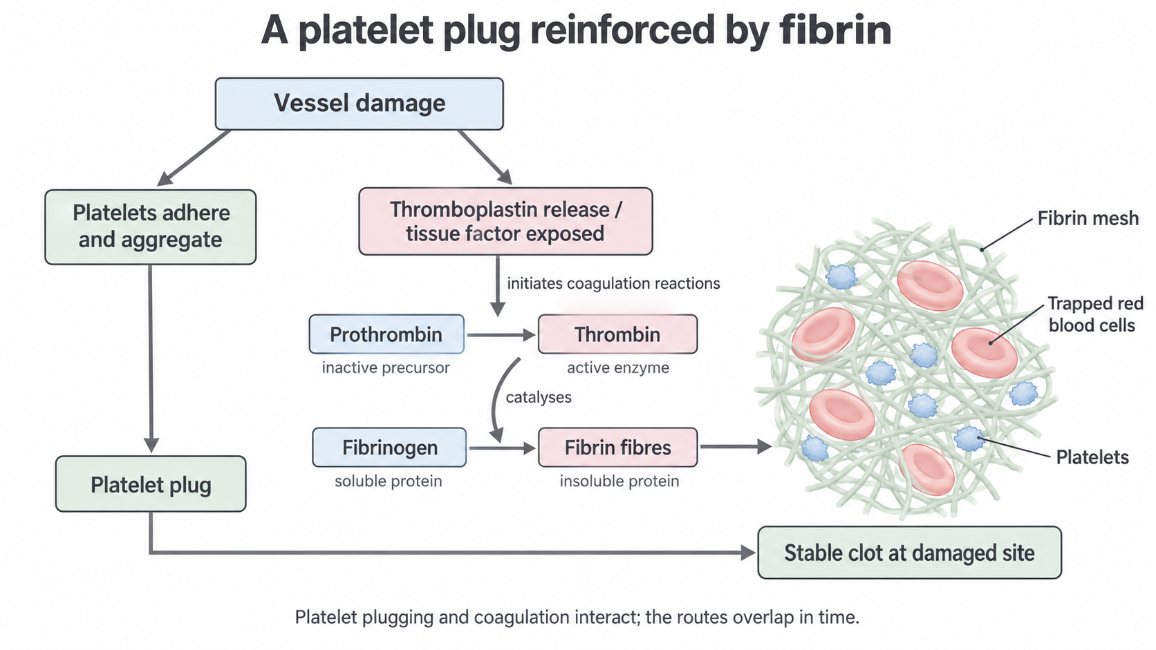
The two protein conversions produce fibrin fibres that reinforce a platelet plug and trap blood cells. The simplified routes overlap in time; thrombin acts on fibrinogen rather than becoming fibrin.
The clot seals the break, reducing further blood loss and providing a temporary support while the vessel repairs. Normal control mechanisms limit clotting to the damaged region, and fibrin is later broken down as repair progresses. The aim is to maintain the seal long enough for healing while restoring blood flow through the repaired vessel.
Predicting a disrupted step
Consider these original, simplified observations from two blood samples after the same clotting trigger. Other relevant conditions are unchanged.
| Sample | Platelet aggregation | Thrombin activity | Fibrin mesh |
|---|---|---|---|
| A | Present | Present | Present |
| B | Present | Blocked | Absent |
Sample B shows why a platelet plug and a fibrin clot must be distinguished. Platelets can aggregate, but blocked thrombin activity prevents the usual fibrinogen-to-fibrin conversion. Therefore, the plug lacks its fibrin reinforcement. The observations do not show that B has no platelets or no fibrinogen; an enzyme can be blocked even when its substrate is available.
Thrombosis and oxygen supply
A clot can become harmful if it grows inside a blood vessel and obstructs the blood supply to tissue beyond it.
Thrombosis
The formation of a blood clot, called a thrombus, within a blood vessel, where it can restrict blood flow.
In the preceding lesson, atherosclerosis produced plaques in artery walls. If a plaque's surface ruptures, blood meets the exposed material beneath the endothelium, including tissue factor. Platelets adhere and aggregate, and coagulation produces thrombin and then fibrin. A thrombus can grow on the damaged plaque. This is a response to injury inside an artery, even though blood is not escaping through a cut in the skin. Research identifying tissue factor in human plaques helps explain why exposed plaque material can initiate clotting.
The plaque and the thrombus are different structures. The plaque forms in the artery wall through atherosclerosis; the thrombus is a blood clot that may form on its damaged surface and extend into the lumen, the space through which blood flows. A thrombus may partly narrow the lumen or completely block it. Thus, a plaque that was not itself closing the artery can become the site of a much more sudden obstruction.
Less blood passing the obstruction means less oxygen delivered to the tissue it supplies. Oxygen is needed for aerobic respiration, which supplies ATP, the cell's immediate energy-transfer molecule, for cell processes. If oxygen shortage is severe and prolonged, cells cannot maintain their normal processes and may die. This chain concerns the tissue downstream of the blocked artery, not simply the blood at the clot.
Coronary and brain arteries
Coronary arteries supply the heart muscle itself. A thrombus in a coronary artery can reduce or stop its blood flow. The resulting oxygen shortage reduces aerobic respiration in the affected cardiac muscle; sustained deprivation can kill that tissue, causing a myocardial infarction, or heart attack. A heart chamber containing oxygenated blood does not compensate for a blocked coronary supply to its muscle. NHLBI's heart-attack explanation links plaque rupture, clot formation and cardiac tissue death.
A clot blocking an artery supplying the brain can similarly deprive brain tissue of oxygen and cause an ischaemic stroke. Some clots form at the eventual obstruction; a fragment of another clot can also detach, travel in the blood and lodge in a narrower artery. Such a travelling fragment is an embolus. This clot-related explanation is for ischaemic stroke: a haemorrhagic stroke instead involves bleeding from a damaged vessel. NHLBI distinguishes these stroke mechanisms.
Clotting is protective when a local clot seals a break during healing. The same platelet and fibrin response contributes to CVD when a thrombus obstructs arterial blood flow, reducing oxygen delivery and threatening the tissue supplied by that artery.
The consequence depends on the obstruction's location, extent and duration. A small local repair clot and a persistent clot blocking a coronary artery use the same clotting chemistry but have very different effects on circulation.