3.4 - Prokaryotic cell ultrastructure
Bacteria carry out the processes of life without a nucleus or mitochondria. Learn how their structures support those processes, how to compare them with eukaryotic cells, and why one historical feature in the specification needs a different interpretation today.
The prokaryotic cell plan
A prokaryotic cell contains genetic information, cytoplasm, ribosomes and a cell-surface membrane, but its DNA is not enclosed in a nucleus.
Prokaryotic cell
A cell without a membrane-bound nucleus. Its main DNA occupies a region of the cytoplasm rather than a compartment enclosed by a nuclear envelope.
Bacteria and archaea are prokaryotes. The familiar model below represents a bacterium; some bacterial features, such as a peptidoglycan wall, must not be assumed to occur in archaea. Ultrastructure means the fine structural detail of a cell. A useful model shows the position and relationship of structures, but it is not a photograph of a particular cell.
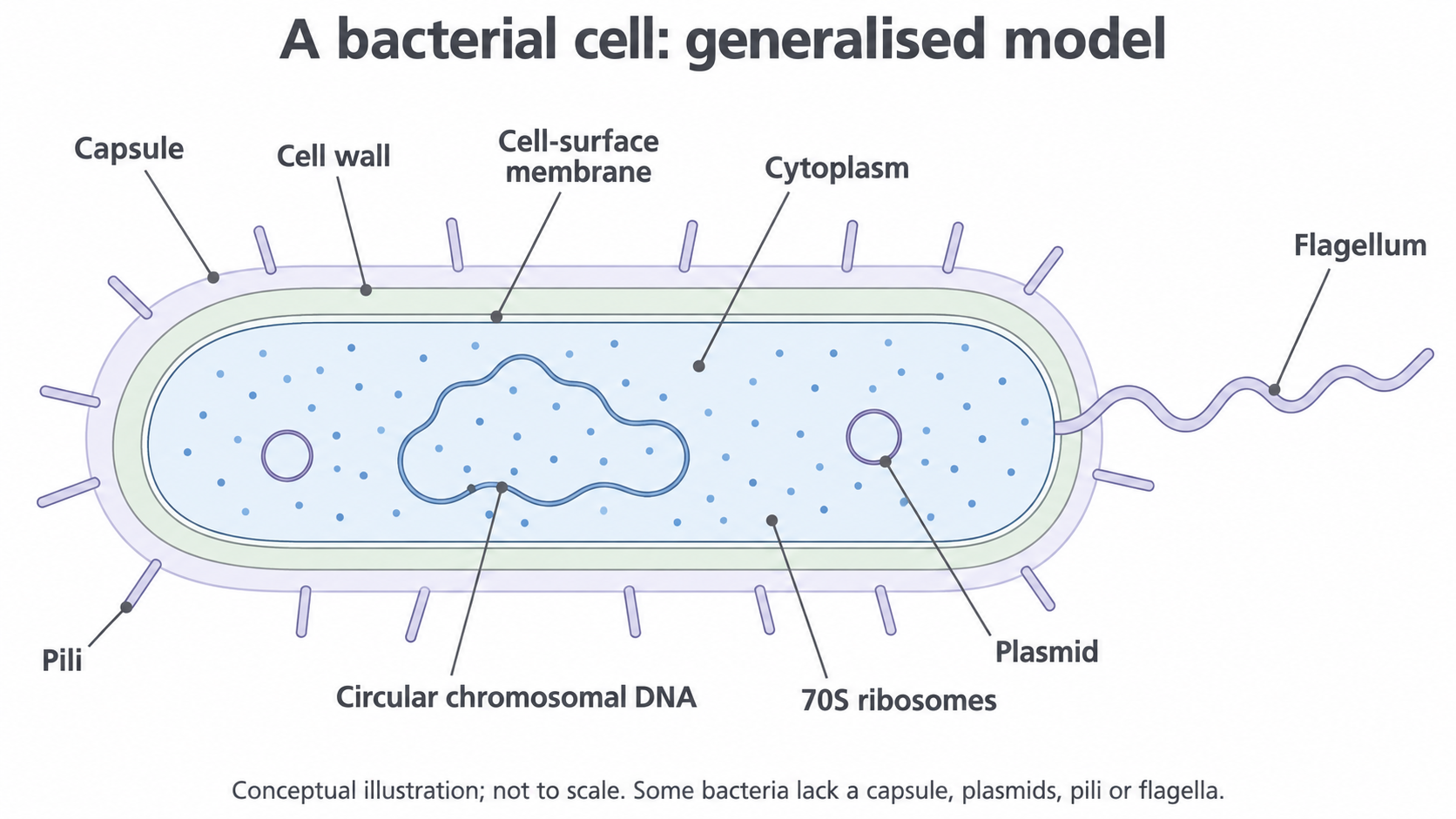
A conceptual bacterial cell model, not an electron micrograph or a scale drawing. Capsule, plasmids, pili and flagella vary between bacteria; they are not present in every bacterial cell. Mesosomes are considered separately as historical preparation artefacts.
The cell-surface membrane separates the cytoplasm from the surroundings and controls movement of substances into and out of the cell. The cytoplasm contains enzymes and is the site of many metabolic reactions. Ribosomes make proteins. These are shared cellular features: having ribosomes or DNA alone does not distinguish a bacterium from a eukaryotic cell.
The major difference is compartmentalisation. A typical eukaryotic cell has a nucleus and membrane-bound organelles such as mitochondria, endoplasmic reticulum and Golgi apparatus. A bacterial cell lacks these structures. Its functions still occur, using different organisation: for example, proteins involved in aerobic respiration are located in the bacterial cell-surface membrane rather than in mitochondria. Absence of mitochondria does not mean absence of respiration.
Use a combination of features to identify a cell. A cell wall alone is insufficient, because plant and fungal cells also have walls. Likewise, one absent organelle is weak evidence: specialised eukaryotic cells can lose particular organelles. DNA lying outside a nucleus together with the absence of membrane-bound organelles is a much stronger comparison.
Cell wall, capsule and membrane
The bacterial cell wall lies outside the cell-surface membrane, while a capsule, when present, forms an additional outer covering.
The cell wall provides mechanical support and helps maintain cell shape. In most bacteria it contains peptidoglycan, also called murein: a strong mesh of carbohydrate chains linked by short peptides. It is chemically different from the cellulose wall of a plant cell. Not every bacterium has a wall, so the diagram is a general model rather than a universal checklist.
Water can enter a bacterial cell by osmosis when its surroundings are sufficiently dilute. The wall resists the resulting expansion and helps prevent the cell from bursting. It does not replace the selectively permeable membrane: the wall provides support, while the membrane regulates exchange. If the wall is weakened, the membrane can remain present yet the cell becomes more vulnerable to bursting in dilute conditions.
A capsule is a relatively organised protective layer outside the wall, commonly made of polysaccharides. It can help a bacterium adhere to a surface and reduce drying. In some pathogenic bacteria it also makes engulfment by phagocytes more difficult, helping the bacterium persist in a host. These benefits depend on the organism and conditions; a capsule does not make every bacterium pathogenic or immune to every defence.
The order from inside to outside is therefore cytoplasm → cell-surface membrane → cell wall → capsule, where all these layers are present. The capsule is not the structure that provides the wall's main resistance to osmotic bursting.
Circular DNA, plasmids and ribosomes
The main bacterial chromosome contains genes needed for cell functions, while plasmids can carry additional genes on separate DNA molecules.
In the typical bacterial model, the main chromosome is one long circular DNA molecule. Circular describes a closed molecule, not a perfectly round shape: it is folded and compacted within a region called the nucleoid. No nuclear envelope surrounds this region. The model is useful at this level, but bacterial chromosome number and shape have exceptions.
Plasmid
A DNA molecule separate from the main chromosome that can replicate independently of it; bacterial plasmids are commonly small circular DNA molecules.
Plasmids may carry genes that give an advantage in particular conditions, such as resistance to an antibiotic. A plasmid is not a small nucleus, a protein or a membrane-bound organelle. It contains genetic instructions; the proteins encoded by those instructions produce the effect. A resistance plasmid may help a bacterium survive a particular antibiotic, but its presence does not imply resistance to all antibiotics. Many bacteria lack a particular plasmid and some have none.
Ribosomes are small structures made of ribosomal RNA and proteins. They assemble amino acids into polypeptides during translation. Bacterial ribosomes are 70S, smaller than the 80S ribosomes in the eukaryotic cytoplasm. Bacteria have no rough endoplasmic reticulum; their ribosomes are in the cytoplasm. The 70S/80S labels identify ribosome types, rather than lengths measured in nanometres. Mitochondria and chloroplasts contain their own bacterial-like ribosomes, so compare bacterial ribosomes with eukaryotic cytoplasmic ribosomes specifically.
| Feature | Typical bacterial cell | Typical eukaryotic cell |
|---|---|---|
| Main DNA location | Nucleoid region of cytoplasm, no nuclear envelope | Inside a membrane-bound nucleus |
| Main chromosome form | Usually circular | Nuclear chromosomes are linear |
| Cytoplasmic ribosomes | 70S | 80S |
| Membrane-bound organelles | No nucleus, mitochondria, ER or Golgi apparatus | Usually present, depending on cell type |
Continue this lesson.
You’re previewing NovaLearn without an account. Create a free account to read the rest.
Create free account