7.2 - The sliding filament mechanism
A skeletal muscle can pull because many tiny contractile units generate force together. This lesson explains how actin and myosin slide past one another, how calcium controls their interaction, and why ATP is needed for both repeated contraction and relaxation.
Sliding within a sarcomere
Inside a muscle fibre, long myofibrils contain repeating units called sarcomeres. This arrangement turns many small molecular movements into shortening along the fibre.
Sarcomere
The repeating contractile unit of a myofibril, extending from one Z line to the next.
Actin forms thin filaments, anchored at the Z lines and extending towards the centre of a sarcomere. Myosin forms thick filaments centred around the M line. Each thick filament has projecting myosin heads that can attach to sites on actin. The thin and thick filaments already overlap in a resting sarcomere; there must be overlap for heads to reach actin.
During shortening, myosin heads pull the actin filaments towards the centre from both ends. Because actin is anchored to the Z lines, those lines move closer together. The sarcomere becomes shorter as overlap increases. Neither the actin filaments nor the myosin filaments become shorter. Sliding changes their relative positions.
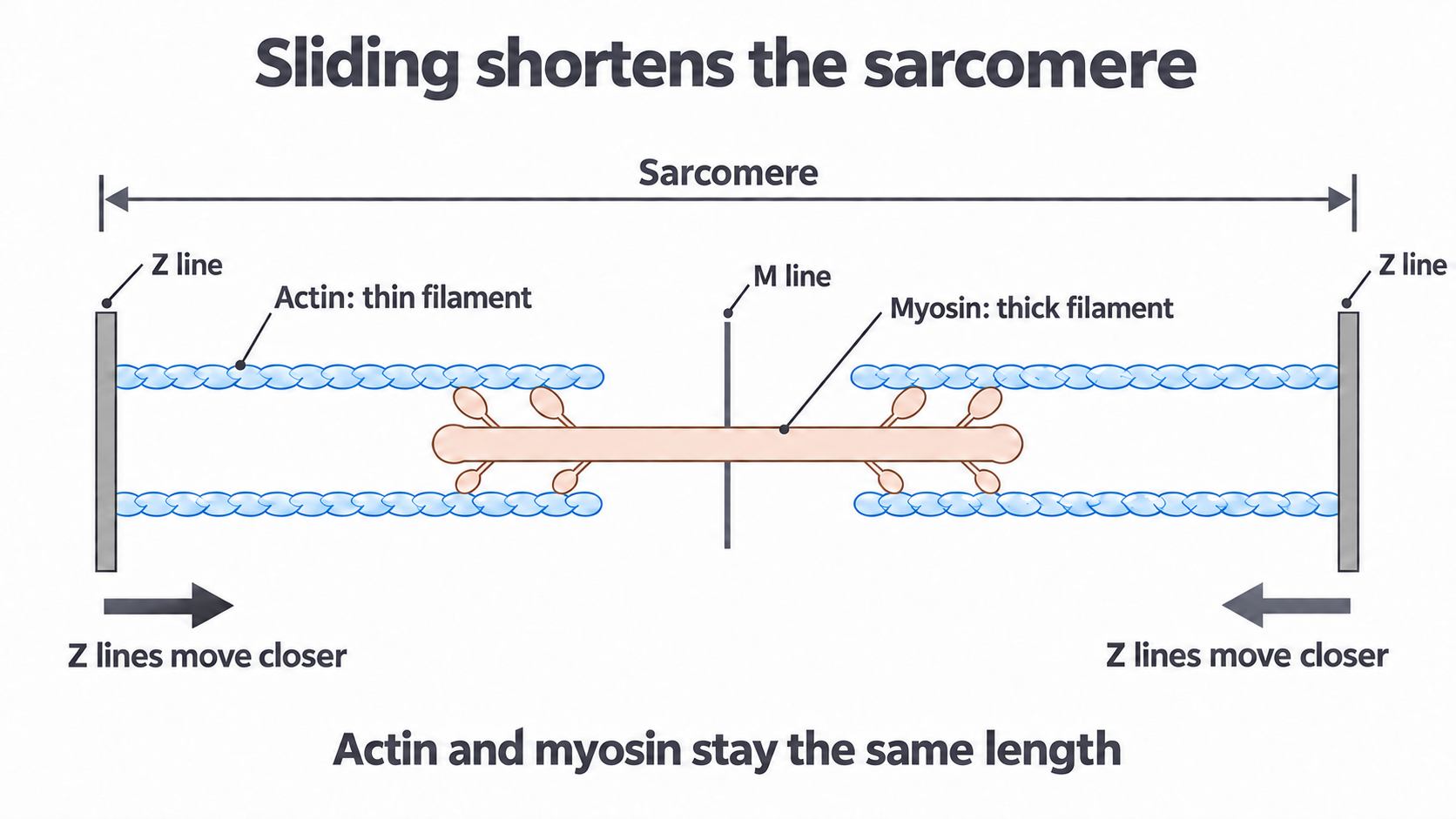
The arrows show the direction in which the Z lines move during shortening. The schematic shows only a few filaments; their individual lengths remain unchanged.
The regular filament arrangement explains the bands seen in striated muscle:
| Region | What occupies it | Change during normal sarcomere shortening |
|---|---|---|
| A band | The full length of the thick myosin filaments, including areas where actin overlaps | Width stays the same because myosin filament length stays the same |
| I band | Thin actin filaments without thick myosin; the Z line lies within it | Becomes narrower as actin slides further into the A band |
| H zone | The central part containing thick myosin without thin actin | Becomes narrower and may disappear as actin approaches the centre |
Sarcomeres are arranged in series along myofibrils, so shortening across many sarcomeres shortens the myofibril and can shorten the whole fibre. Force also depends on the load: an active muscle can develop tension while its overall length stays constant.
Calcium exposes actin sites
The availability of binding sites on actin controls whether myosin heads can form force-producing attachments. Two proteins associated with the thin filament provide this control: tropomyosin, a long protein lying along actin, and troponin, a protein complex associated with tropomyosin.
In a resting fibre, the calcium ion concentration in the sarcoplasm is low. The sarcoplasm is the cytoplasm of a muscle fibre. Tropomyosin lies in a position that blocks myosin-binding sites on actin. Myosin and actin can therefore be close together without repeated force-producing attachments.
Stimulation of a skeletal muscle fibre causes its sarcoplasmic reticulum, an internal membrane system that stores calcium, to release into the sarcoplasm. The next events form a causal sequence:
- Calcium ions bind to troponin.
- Troponin changes shape, moving the associated tropomyosin away from the myosin-binding sites on actin.
- Myosin heads can now attach to those exposed sites and generate force through the cycle described next.
Calcium is a regulatory signal: it permits the interaction. ATP supplies the chemical energy used by the molecular motor. Calcium does not pull the actin filament or replace ATP. Likewise, calcium binds to troponin in this switch; it does not bind to tropomyosin to make tropomyosin contract.
This mechanism predicts that increasing sarcoplasmic calcium will fail to activate a filament if its troponin cannot respond to calcium. A signal can be present while the protein that receives it cannot transmit its effect.
ATP and the cross-bridge cycle
A myosin head repeatedly attaches, pulls, detaches and resets while calcium keeps the actin sites accessible. An attachment between a myosin head and actin is a cross-bridge. Its temporary nature allows the head to reach a new site after each pull.
ATPase
An enzyme that catalyses ATP hydrolysis. The myosin head has ATPase activity, converting ATP and water to ADP and inorganic phosphate.
ATP hydrolysis is coupled to changes in the shape of the head. The net hydrolysis reaction can supply usable energy; breaking a bond alone requires energy and is not, by itself, an explanation for energy release. In the following cycle, means inorganic phosphate.
Detach, reset, attach and pull
There is no unique first step in a cycle. Start with a head that has finished a pull and is still attached to actin:
Continue this lesson.
You’re previewing NovaLearn without an account. Create a free account to read the rest.
Create free account