2.5 - Nucleotides, DNA and RNA
DNA and RNA are built from small units called nucleotides. Their sugars, bases and bonds explain both how a strand is held together and how two DNA strands fit together. Learning these structures gives us a foundation for understanding how cells use genetic information.
The three components of a nucleotide
A nucleotide is one building unit of a nucleic acid, such as DNA or RNA.
Mononucleotide
A unit containing a pentose sugar linked to a phosphate group and a nitrogen-containing base.
Pentose means a sugar with five carbon atoms. DNA nucleotides contain deoxyribose; RNA nucleotides contain ribose. Both sugars have five carbons. Deoxyribose has one fewer oxygen atom: at its 2′ carbon it has H where ribose has an OH group. The prime symbol in 2′, pronounced “two prime”, distinguishes a sugar position from positions in the base.
The base attaches to the sugar's 1′ carbon. In the mononucleotide shown below, the phosphate is linked through an oxygen to the sugar's 5′ carbon. The phosphate and base therefore attach to the sugar, rather than the phosphate attaching directly to the base. The sugar also has an OH group at 3′, which matters when nucleotides join into a strand.
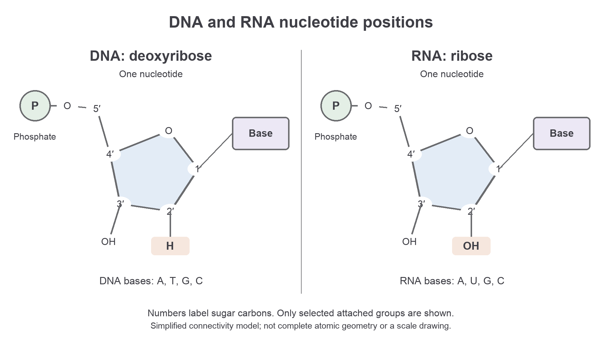
Simplified nucleotide structures. The ring contains four carbons and one oxygen; the fifth carbon lies outside the ring. Only the groups needed to compare the nucleotides are shown, rather than every atom or three-dimensional bond angle.
The nitrogen-containing bases are adenine (A), guanine (G), cytosine (C), thymine (T) and uracil (U). DNA uses A, G, C and T. RNA uses A, G, C and U. A nucleotide contains one base, not all four bases used by its nucleic acid. For example, an adenine DNA nucleotide contains adenine, deoxyribose and phosphate.
Joining nucleotides into a strand
A nucleic acid strand is a polymer made from many nucleotide units joined in a chain.
Polynucleotide
A chain of nucleotide units joined by covalent phosphodiester bonds in a sugar–phosphate backbone.
Nucleotides join through condensation reactions. In the simple condensation model, an H and an OH are removed as water while a covalent linkage forms between the phosphate group of one nucleotide and the sugar of another. The resulting phosphodiester linkage connects the 3′ position of one sugar, through oxygen and phosphate, to the 5′ position of the next sugar. This is the linkage to recognise in both DNA and RNA. Cellular enzymes use activated nucleotides to build strands; the simple condensation model describes the structural linkage, rather than all the chemical steps of enzyme-catalysed synthesis.
Repeated joining produces an alternating sugar–phosphate backbone. Bases project from the sugars and are not the links that join successive nucleotides along a strand. A phosphodiester bond is covalent: it is different from the hydrogen bonds between paired bases that we will meet next. Breaking the backbone would break a strand into shorter chains; separating two paired strands can leave both backbones intact.
The chemical arrangement gives a strand two different ends, called 5′ and 3′. These names refer to sugar-carbon positions, not a count of bases. A linear chain with four nucleotides needs three links between successive nucleotide units. More generally, a linear chain with nucleotide units has links between them; this count excludes a connection joining the two ends into a circle.
Within a DNA or RNA strand, covalent phosphodiester bonds join nucleotide units through the sugar–phosphate backbone. Hydrogen bonds between bases do a different job.
The distinction lets us reason about a damaged molecule without treating every bond in it as interchangeable.
Complementary strands form the double helix
The usual DNA molecule consists of two complementary polynucleotide strands twisted around each other.
Complementary base pairing means that a base has a specific matching partner: adenine pairs with thymine, and guanine pairs with cytosine. The shapes and arrangements of hydrogen-bonding groups on the bases allow these pairs to fit together. In ordinary DNA base pairs, A–T forms two hydrogen bonds and G–C forms three hydrogen bonds.
The bases point inward, towards the bases on the other strand. Hydrogen bonds form between the paired bases on opposite strands, helping to hold those strands together. Covalent phosphodiester bonds join nucleotide units along each backbone; they do not run across the paired bases. Each hydrogen bond is relatively weak compared with a covalent backbone bond, but many base pairs together stabilise the paired structure. Base-stacking interactions also contribute to stability, so hydrogen-bond number alone is not a complete explanation of every difference in DNA stability.
The two strands run in opposite directions: beside the 5′ end of one is the 3′ end of the other. This arrangement is called antiparallel. Their sugar–phosphate backbones lie on the outside, while the complementary base pairs lie inside. The whole paired structure twists into a double helix: two strands winding around the same axis.
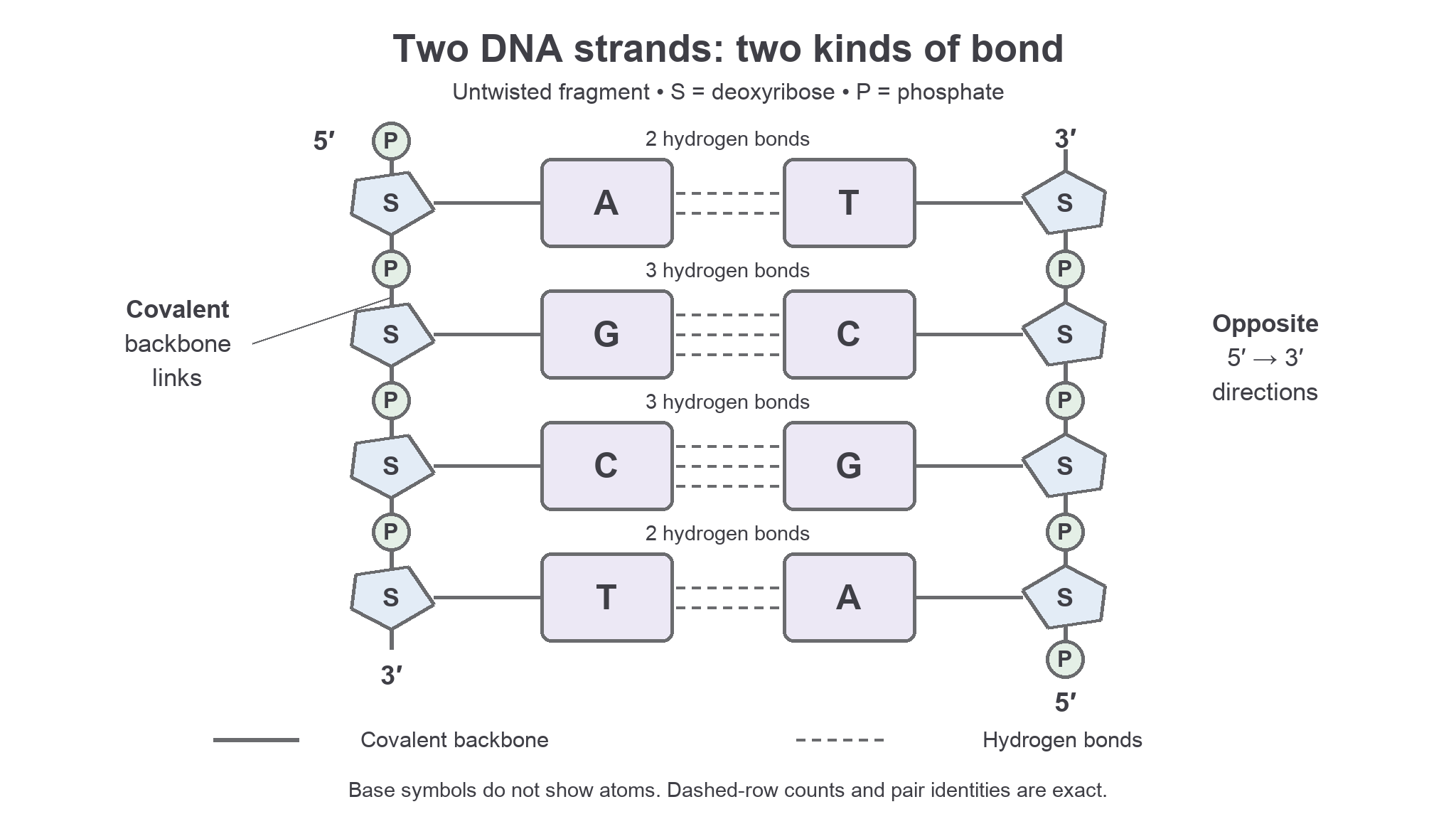
An untwisted DNA fragment makes the bond types visible. Solid backbone links connect nucleotide units along each strand; dashed links represent hydrogen bonds across each base pair. The strands run 5′ to 3′ in opposite directions.
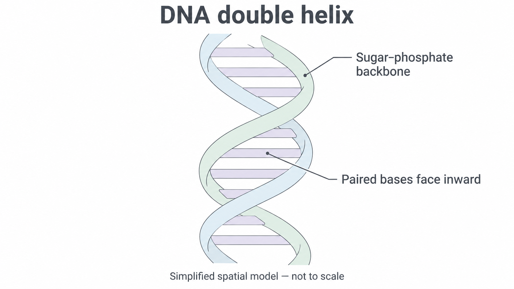
The same two-strand arrangement twists into a double helix. This spatial model does not show individual atoms or hydrogen-bond counts.
The pairing rule explains why two strands are complementary rather than identical. Opposite A is T, not another A. It also explains why separation of paired strands need not destroy their covalent backbones.
Using the base-pairing rule
Knowing the bases on one DNA strand lets us determine the matching bases on its partner.
Suppose one strand is written 5′–AGCTTA–3′. Match each position using A–T and G–C, and put the opposite ends beside one another:
| Strand | Left end | Bases, read left to right | Right end |
|---|---|---|---|
| Given strand | 5′ | A G C T T A | 3′ |
| Complementary strand | 3′ | T C G A A T | 5′ |
The complementary strand is 3′–TCGAAT–5′ in this alignment. If asked to write that same complementary strand from its own 5′ end to its 3′ end, read its row in reverse: 5′–TAAGCT–3′. Changing the writing direction changes the order on the page, not the actual pairings. This is a structural sequence task; we do not need to group bases into codons to solve it.
Whole-molecule base proportions
In ordinary double-stranded DNA, every A is paired with a T and every G with a C. Therefore, across both strands together, the number of A bases equals the number of T bases, and the number of G bases equals the number of C bases. The corresponding percentages are equal too. This does not require A and T to occur in equal numbers on one individual strand. A single strand, whether DNA or RNA, need not have a complementary partner for every base within that same strand, so its G and C counts need not be equal either.
For example, if C makes up 24% of all bases in a double-stranded DNA sample, G is also 24%. Together they account for 48%, leaving for A and T. Each of these is . Check that .
Counting bases and hydrogen bonds
A fragment with 10 base pairs contains 20 nucleotide units, because each pair contains one nucleotide from each strand. If four of those pairs are G–C and six are A–T, their total hydrogen-bond count is . Count pairs before multiplying: counting all individual G and C bases as separate G–C pairs would double the answer.
Comparing DNA and RNA structures
DNA and RNA share a nucleotide-based structure, but their differences should be stated as paired comparisons.
| Feature | DNA | RNA |
|---|---|---|
| Sugar in each nucleotide | Deoxyribose; H at 2′ | Ribose; OH at 2′ |
| Normal base set | A, T, G, C | A, U, G, C |
| Usual cellular structure | Two complementary antiparallel strands in a double helix | One polynucleotide strand, which may fold |
| Backbone | Alternating sugar and phosphate joined by covalent phosphodiester bonds | Alternating sugar and phosphate joined by covalent phosphodiester bonds |
Both are nucleic acids, both are polynucleotides, and both contain A, G and C. A statement such as “RNA contains phosphate but DNA contains sugar” is wrong: each contains both, although the sugar differs. Likewise, uracil is a base, not the name of RNA's sugar.
RNA is usually single-stranded in cells, but that does not mean every RNA molecule is a straight chain with no hydrogen bonds. Complementary regions within a single RNA strand can pair as it folds. In ordinary RNA base pairing, A pairs with U and G pairs with C. This local folding does not make the whole molecule two separate DNA strands. Avoid treating “RNA is always single-stranded and cannot base-pair” as a universal rule.
Consider a laboratory description: “Molecule X is a polynucleotide containing ribose and uracil; some regions fold back and pair.” Ribose and uracil support identifying X as RNA. The fold does not overturn that identification. Conversely, observing A, G and C alone would not distinguish DNA from RNA because both contain those bases. Identify a molecule using the features that actually discriminate between them, rather than a feature they share.
Compare like with like: sugar with sugar, base set with base set, and strand arrangement with strand arrangement. Both DNA and RNA have a covalent sugar–phosphate backbone.