1.3 - Arteries, veins and capillaries
Blood vessels must carry blood over long distances and bring its contents close enough to cells for exchange. Learn why arteries, capillaries and veins have different walls and lumens, and how these differences suit the pressures and movements of blood through them.
Vessels and blood direction
Arteries carry blood away from the heart, capillaries provide exchange surfaces, and veins return blood towards the heart.
An artery divides into smaller arteries and then arterioles, small arteries that supply capillary networks. Capillaries merge into venules, small vessels that join to form veins. Along this route, the blood itself moves by mass transport; substances then move between blood and nearby tissues across the capillary wall.
Lumen
The internal space of a blood vessel through which blood flows.
The lumen is different from the surrounding wall. A large lumen does not mean a thick wall. The innermost surface of all three vessel types is lined by endothelium, a single layer of flattened cells. Arteries and veins also have supporting tissue around this lining; capillaries have a much thinner wall.
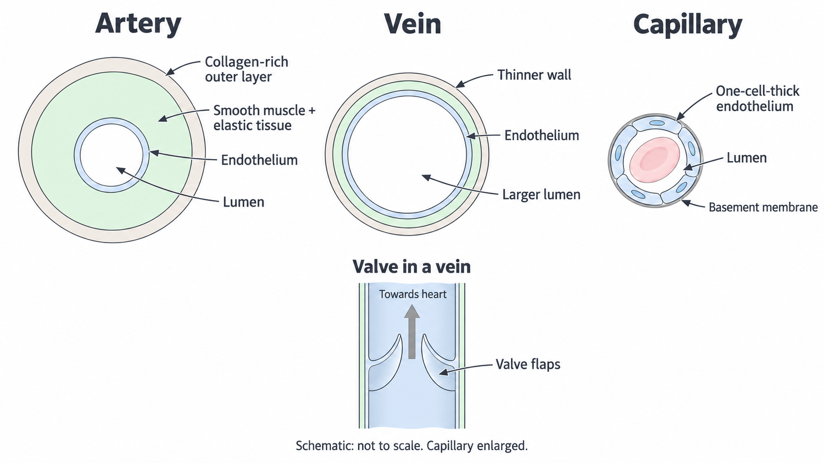
The artery has a thicker wall relative to its lumen than the vein. The enlarged capillary has one layer of endothelial cells and a thin basement membrane. The valve inset shows permitted flow towards the heart; all drawings are schematics, not measured sections.
Compare the drawings by following the same route from lumen to outside: endothelial lining, any muscle and elastic tissue, then outer supporting tissue. The amount of each tissue matters, rather than simply whether a vessel contains it. The capillary is enlarged so that its wall can be seen; a real capillary is far narrower than a typical artery or vein.
The words artery and vein describe direction, rather than oxygen concentration. In the body circuit, arteries usually carry oxygenated blood and veins usually carry blood with less oxygen. In the lung circuit, the pulmonary artery carries blood with less oxygen away from the heart to the lungs, while pulmonary veins return oxygenated blood to the heart. Both circuits obey the direction rule.
Arteries withstand and smooth pressure
Arteries receive blood pushed out by the heart, so their walls must withstand pressure and accommodate repeated surges of blood.
Blood pressure is the force per unit area exerted by blood on its surroundings. A pressure difference drives blood from a region of higher pressure towards a region of lower pressure. Within a circuit, arteries generally experience greater pressure than the veins returning blood from that circuit. Pressure also fluctuates with each heartbeat, most strongly in the large arteries close to the heart.
An artery has a relatively thick wall containing smooth muscle, elastic tissue and strong collagen fibres. Collagen helps resist excessive stretching and prevents the wall from being damaged by the pressure. Elastic tissue can stretch when a surge of blood enters and then recoil, returning towards its original shape as pressure falls between heartbeats. Recoil maintains pressure on the blood and helps sustain forward flow while the heart is not ejecting blood. It also makes the flow less sharply intermittent than it would be through a rigid tube.
Recoil is a passive property of elastic tissue. It is different from active contraction of smooth muscle. Circular smooth muscle in the wall can contract to narrow the lumen or relax to widen it. In the smaller arteries and especially arterioles, this alters resistance to flow and controls how much blood reaches a capillary bed. For the same pressure difference, narrowing a vessel increases resistance and reduces the flow through it. Widening it has the opposite effect.
Large arteries near the heart contain abundant elastic tissue for handling the pulse. More distant arteries have a greater emphasis on muscular control of distribution. The proportions change along the arterial system, so a single drawing represents the general design rather than every artery's exact wall.
Compared with a similar-sized vein, an artery usually has a smaller lumen relative to its wall thickness. This is a useful structural comparison, but the lumen does not create the driving force: the heart establishes the pressure difference. There are no repeated pocket valves along ordinary arteries; valves at the exits of the heart prevent blood returning into the heart.
Capillaries bring blood close to cells
Capillaries are specialised for exchange between blood and the tissue fluid surrounding cells.
A capillary's wall is a single layer of flattened endothelial cells supported by a very thin basement membrane, a non-cellular supporting layer. It has no thick muscular or outer supporting layer like an artery or vein. This gives substances a short diffusion distance across the vessel wall. The tissue fluid is the immediate link between most tissue cells and the blood: substances cross the capillary wall, pass through tissue fluid and then enter or leave cells.
The lumen of a typical capillary is so narrow that red blood cells pass through in single file, often deforming as they do so. This brings their contents close to the exchange wall. Many branching capillaries form an extensive network through a tissue, providing a large total surface area and placing most supplied cells close to a blood vessel. A very thin wall would be less useful if the nearest capillary were far from the cells needing oxygen.
Blood moves relatively slowly through capillary beds, giving time for exchange. Although each capillary has a tiny cross-sectional area, the many capillaries operating in parallel have a very large total cross-sectional area. The incoming blood is distributed among them. This explains the low average speed through the bed; it is misleading to say that slow capillary flow is simply caused by each lumen being narrow. Pressure is also lower than in the supplying arteries, with a substantial pressure drop across the arterioles before blood reaches the capillaries.
Diffusion needs a concentration difference as well as a short distance. In a respiring body tissue, cells use oxygen and nutrients such as glucose, so these can move from blood towards cells. Cells produce carbon dioxide, which can diffuse towards the blood. Continuing blood flow supplies more oxygen and removes carbon dioxide, helping to maintain these differences. In the lungs, the local oxygen and carbon dioxide gradients differ, so gas exchange occurs in the opposite directions. Direction of diffusion follows the local concentration difference, not a rule that everything always leaves a capillary.
Capillary walls are permeable to substances that need to be exchanged. Small dissolved substances can cross through or between endothelial cells, depending on the substance and the capillary. Ordinary tissue capillaries retain red blood cells and most large plasma proteins; their walls are not unrestricted holes. Permeability differs between organs, so do not assume that every capillary has identical gaps or pores.
Veins return blood at low pressure
Veins collect blood after it has passed through capillary beds and return it towards the heart under a much smaller pressure difference.
Compared with a similar-sized artery, a vein has a thinner wall, with less smooth muscle and elastic tissue, and a larger lumen. The lower pressure makes a thick pressure-resistant wall less necessary. A wide lumen offers relatively low resistance, allowing blood to return even when the driving pressure difference is small. The wall still contains muscle, elastic tissue and collagen; saying that veins have none of these tissues is incorrect. Their thinner walls are more easily compressed and a vein may look partly collapsed in a prepared tissue section. Wall thickness relative to lumen size is more useful for identification than expecting every vessel to be perfectly circular.
Many veins, especially in the limbs, contain valves formed from folds of the inner lining with supporting connective tissue. They open when blood pushes in the permitted direction and close if blood starts to move backwards. This helps prevent blood flowing back towards the tissues, particularly when return from the legs is against gravity. Not every vein has valves.
Contraction of surrounding skeletal muscles compresses veins and raises pressure in that section of vein. Blood can move towards the heart through the valve ahead of the compressed region; the valve behind closes against reverse flow. As muscles relax, the vein can fill again. Repeated muscle contractions and one-way valves therefore work together to assist venous return. The valves themselves do not contract or add energy to the blood: they prevent backflow.
To compare vessels, choose the same feature for both rather than listing unrelated facts. For example, both arteries and veins have endothelial linings, but arteries have more substantial muscular and elastic walls for higher pressure, whereas veins have thinner walls and wider lumens for low-pressure return. Capillaries retain the thin endothelial exchange wall and dispense with those thick surrounding layers.
| Feature | Arteries | Capillaries | Veins |
|---|---|---|---|
| Main role | Carry blood away from heart and distribute it | Exchange between blood and tissues | Return blood towards heart |
| Pressure within a circuit | Relatively high and pulsatile, especially near heart | Lower after the arterioles | Relatively low |
| Wall | Thick supporting, muscular and elastic tissue around endothelium | One endothelial cell layer with thin basement membrane | Thinner supporting, muscular and elastic tissue around endothelium |
| Lumen | Smaller relative to wall than a comparable vein | Very narrow individually; large total area of a network | Larger relative to wall than a comparable artery |
| Valves along the vessel | Absent in ordinary arteries | Absent | Present in many veins, especially limbs |
Link each feature to its job: an artery handles pressure and distributes blood, a capillary minimises exchange distances, and a vein supports low-pressure return with a wide lumen and, where present, one-way valves.
Use this relationship to predict changes. For example, compressing a vein need not stop venous return: intermittent compression by contracting skeletal muscles can help it, provided the valves direct the resulting movement towards the heart.