2.12, 2.14 - Mutations and cystic fibrosis
In 2.12, you will see how a copying error can change DNA and why several different mutations can cause cystic fibrosis. In 2.14, you will follow the consequences from a membrane protein to gas exchange, digestion and reproduction. The same molecular fault can affect several organs because their epithelial cells use the same protein for different fluid-transport tasks.
How copying errors become mutations
DNA replication usually produces an accurate copy, but an uncorrected error can leave a lasting change in the base sequence.
Gene mutation
A gene mutation is a change in the DNA base sequence of a gene. It can produce a new allele, which is a different version of that gene.
During replication, the two DNA strands separate and each acts as a template. DNA polymerase joins nucleotides to form a new strand, normally using complementary pairing: A with T, and C with G. Occasionally an incorrect nucleotide is incorporated. For example, a template G may be paired with T rather than C. Proofreading and repair correct many errors. If the mismatch escapes correction and DNA is copied again, one descendant DNA molecule can contain a changed, fully paired base sequence. The mistake is then a mutation rather than a temporary mismatch.
Copying can also add or miss nucleotides, for example if the strands slip out of alignment. A substitution replaces a nucleotide; an insertion adds one or more nucleotides; a deletion removes one or more. In double-stranded DNA, the corresponding change becomes established in the complementary strand too. These names describe what happened to the sequence, not how serious its effect will be.
Mutations arise spontaneously; some mutagens, such as particular chemicals or forms of radiation, increase their frequency by damaging DNA or interfering with accurate copying. A mutation does not arise because a cell needs a particular improvement. A change in a body cell can be passed to its daughter cells, whereas transmission to offspring requires the change to be present in the genetic material of a gamete. A person with an inherited CF-causing allele did not necessarily acquire a new copying error during their own development.
From a base change to a protein change
A mutation can change a protein because transcription copies the DNA information into mRNA and translation reads that mRNA in groups of three bases.
The reading frame is the grouping of bases into consecutive triplets from the translation start. A DNA coding strand has the same sequence as the mRNA, read 5′ to 3′, except that DNA uses T and RNA uses U. The complementary template strand is read in the opposite direction during transcription. A stop codon ends translation; it does not add an amino acid.
The following complete, short coding sequence is an original teaching example, not part of the CFTR gene. Spaces show the reading frame, not gaps in the molecule.
| Molecule, with direction | Sequence or translation |
|---|---|
| DNA coding strand, 5′ to 3′ | 5′-ATG GAA TTT CCG TAA-3′ |
| DNA template strand, 3′ to 5′ | 3′-TAC CTT AAA GGC ATT-5′ |
| mRNA, 5′ to 3′ | 5′-AUG GAA UUU CCG UAA-3′ |
| Translation, from the start codon | Methionine–glutamate–phenylalanine–proline, then stop |
These mRNA codons supply every translation used in this part: AUG = methionine; GAA and GAG = glutamate; UUU and UUC = phenylalanine; UAU = tyrosine; CCG = proline; GAU = aspartate; CGU = arginine; UAA = stop.
Substitutions and the genetic code
Changing the coding-strand triplet GAA to GAG changes the mRNA codon too, but both code for glutamate. This is a synonymous mutation: the amino acid sequence is unchanged in this example because the genetic code is degenerate. Changing TTT to TAT gives UAU in mRNA and replaces phenylalanine with tyrosine. This is a missense mutation, which changes an amino acid. Changing GAA to TAA gives UAA and ends translation after the first amino acid: a nonsense mutation introduces a premature stop codon.
Different amino acids have different side chains, or R groups. A replacement may alter the interactions between these groups that hold a protein in its three-dimensional shape, changing an active site, a channel gate or protein stability. Its effect depends on the position and the amino acids involved. Some replacements have little effect; others strongly reduce function. A premature stop may prevent a full-length functional protein being made. Mutations outside protein-coding triplets can also affect the amount of protein produced or the processing of its RNA. A DNA change therefore does not automatically mean a harmful amino acid change.
Deletions and the reading frame
Deleting the whole TTT triplet gives coding DNA 5′-ATG GAA CCG TAA-3′ and mRNA 5′-AUG GAA CCG UAA-3′. The protein loses phenylalanine, but the downstream triplets remain in their original frame. A deletion of three bases, or another multiple of three, is in-frame within a coding region; the protein can still be impaired even though the frame is preserved.
Instead, delete the first A in the original GAA triplet. The remaining coding-strand fragment regrouped from the same start is 5′-ATG GAT TTC CGT AA-3′. Its mRNA is 5′-AUG GAU UUC CGU AA-3′. The complete codons shown now give methionine–aspartate–phenylalanine–arginine. The final AA is an incomplete codon, so this fragment alone does not specify the next amino acid or a new stop position. This frameshift changes the downstream grouping. Insertions or deletions not divisible by three cause a frameshift within a coding region, often altering many amino acids and sometimes introducing an early stop.
Mutation type does not give a universal severity ranking. An in-frame deletion can destroy an important function, while a substitution can be synonymous. Predict the consequence from the particular sequence and protein role.
The way to analyse an unfamiliar sequence is to identify the strand and direction, make or read the mRNA, group codons from the stated start, then use the supplied genetic code. Compare the amino acid sequence and the reading frame separately.
Different mutations can impair CFTR
Cystic fibrosis can result from several different mutations in the gene that encodes CFTR, rather than from one unique DNA error.
CFTR, the cystic fibrosis transmembrane conductance regulator, is a membrane protein that forms a regulated channel for chloride ions. It also conducts bicarbonate ions. Its function depends both on reaching the appropriate epithelial cell membrane and on working when it is there. In cystic fibrosis, CF-causing changes in both copies of the gene reduce effective CFTR function. The two copies may carry the same mutation or different mutations; inheritance patterns are developed in 2.13.
A well-known example is F508del, also written ΔF508. Three DNA bases are deleted, leading to the loss of phenylalanine at amino acid position 508. This is an in-frame deletion, not a one-base frameshift. The altered protein folds and is processed poorly, so much of it is removed by the cell before reaching the surface. Less functional CFTR is then available in the membrane. The short fictional sequence in the previous part illustrates an in-frame deletion; it is not the actual F508del sequence.
Another CF-causing mutation, G551D, substitutes aspartate for glycine at position 551. CFTR can reach the membrane but its channel opens poorly: a gating defect. Other mutations introduce premature stop codons, interfere with RNA splicing or reduce channel conduction or protein quantity. Different DNA changes can therefore converge on reduced chloride transport through different mechanisms. A protein can be present but ineffective, or effective protein can be largely absent.
Consider this original, qualitative example of cells engineered to make particular CFTR variants. All rows are compared under the same channel-activating conditions; the labels are illustrative observations, not clinical measurements.
| CFTR produced by the cells | Full-length CFTR detected at the cell surface | Chloride transport response |
|---|---|---|
| Unchanged reference CFTR | Abundant | Large |
| F508del CFTR | Very little | Small |
| G551D CFTR | Present at an amount similar to the reference | Small |
For F508del, the small transport response is consistent with little protein reaching the surface. For G551D, a similar surface amount but a smaller response supports impaired transport by the surface protein, rather than reduced quantity alone. Detecting CFTR therefore cannot establish normal channel function. The table separates location from transport; it does not give an exact estimate of a person's lung function or fertility. Residual CFTR function, other genes, environment, infections and treatment all influence the phenotype. People with cystic fibrosis do not necessarily have identical symptoms or severity.
CFTR, water and mucus hydration
CFTR changes the movement of ions across an epithelium, which in turn changes the movement of water and the properties of its secretions.
An epithelium is a layer of cells lining a surface or cavity. The lumen is the space inside a tube or cavity, such as an airway or pancreatic duct. In secretory airway epithelia, CFTR in the membrane facing the lumen provides a route for chloride ions, , to move into the airway fluid. The ions pass through a channel down their electrochemical gradient; CFTR is not a pump carrying water. Other membrane transport systems help establish the ion gradients and regulate fluid absorption.
Ion movement affects the dissolved-solute concentration on either side of an epithelium. Water moves by osmosis towards the region with fewer free water molecules. In a simplified secretory model, chloride secretion helps water move into the lumen and maintains a hydrated surface fluid. CFTR also contributes to bicarbonate transport and the conditions in which mucus unfolds and functions. The useful overall connection is effective ion transport → suitable fluid hydration → mobile mucus.
When effective CFTR function is reduced, normal fluid balance is disrupted. Airway surface fluid can become depleted and mucus becomes less hydrated and more viscous: it resists movement. Water moves through the epithelium by its own routes, including water-permeable membranes and spaces between cells, rather than through the CFTR chloride channel.
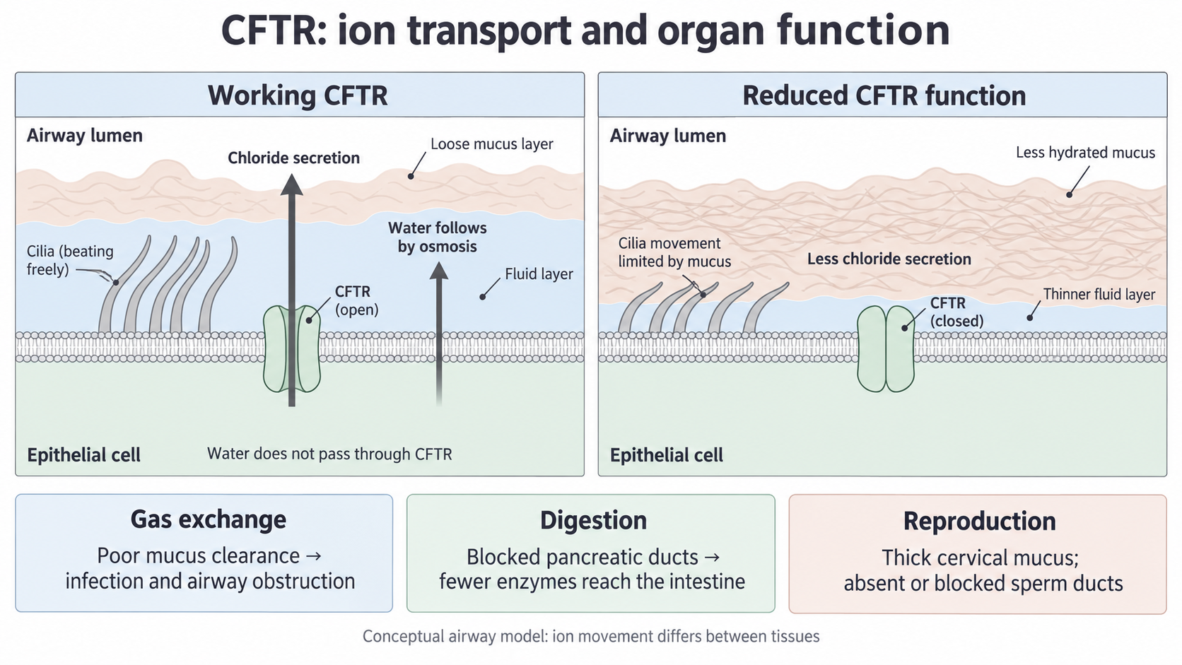
The schematic follows a secretory airway model. The closed-channel symbol illustrates reduced effective transport; some CF mutations instead leave little CFTR at the membrane. The lower boxes preview the organ consequences developed below. The picture is conceptual, without a scale or exact molecular topology.
The direction of chloride movement depends on the tissue and its gradients. In sweat ducts, CFTR supports chloride reabsorption from the duct fluid rather than airway-style secretion. Reduced reabsorption leaves more salt in the final sweat. Do not extend the airway arrow into a claim that CFTR always moves chloride out of cells, or that cystic fibrosis lowers salt in every secretion. Sweat physiology is a useful check on the model; screening decisions belong to 2.15–2.16.
Why gas exchange is impaired
In the respiratory system, thick secretions interfere with both airway clearance and delivery of fresh air to gas-exchange surfaces.
Normally mucus traps particles and microorganisms in the conducting airways. Cilia are small beating projections on epithelial cells that move the mucus towards the throat, where it can be swallowed or expelled. An adequately hydrated fluid layer lets this transport work. These conducting-airway cells should not be confused with the thin alveolar exchange surface.
With reduced CFTR function, dehydrated, viscous mucus is harder to move. Ciliary clearance becomes less effective, so microorganisms can remain in the airways and multiply. Recurrent infection stimulates inflammation and further secretions. Infection and inflammation can damage lung tissue; persistent damage can reduce effective exchange area or increase the barrier that gases must cross. This gives a cycle in which poor clearance encourages infection, which causes further tissue impairment.
Mucus can also obstruct small airways. Less fresh air reaches the alveoli supplied by those airways, so oxygen concentration in their air can fall and carbon dioxide can accumulate. The concentration difference driving oxygen diffusion into the blood is then reduced; carbon dioxide removal is also impaired. In affected regions, reduced ventilation and tissue damage can together lower the rate of gas exchange. This can reduce oxygen availability for aerobic respiration and contribute to breathlessness and reduced exercise tolerance.
The mechanism is not that a CFTR channel normally transports oxygen or carbon dioxide. CFTR transports ions; its effect on fluid and mucus changes airway and lung function, which changes gas diffusion. Nor does mucus simply coat every alveolus in the same way. Begin with the affected airway or exchange barrier and follow the consequence relevant to that location.
Why digestion and absorption are impaired
In the digestive system, abnormal secretions can prevent pancreatic enzymes from reaching the food they must digest.
The pancreas produces digestive enzymes and releases them through ducts into the first part of the small intestine. Proteases digest proteins, amylase digests starch and lipase digests triglycerides. Digestion converts large food molecules into smaller molecules that can be absorbed through the intestinal epithelium. CFTR in duct epithelial cells contributes to chloride, bicarbonate and fluid secretion; bicarbonate also helps neutralise acidic material entering from the stomach and provides suitable conditions for intestinal digestion.
Reduced CFTR function can make pancreatic secretions thick and difficult to move. They can obstruct the pancreatic ducts, so fewer enzymes reach the small intestine. Continued blockage can damage the enzyme-producing tissue, further reducing enzyme supply: pancreatic insufficiency means the pancreas does not provide enough digestive enzymes for normal digestion.
For example, with too little lipase entering the intestine, less triglyceride is hydrolysed to absorbable products. More fat remains in the intestinal contents and is lost in faeces. Absorption of fat-soluble vitamins can also be impaired. Reduced digestion of other nutrients can contribute to lower nutrient and energy uptake, poor growth or difficulty maintaining body mass. The key distinction is that the first defect in this chain is delivery of enzymes, rather than a mutation directly removing every intestinal absorption protein.
Thick intestinal contents can also obstruct the intestine. The precise digestive phenotype varies: some people retain enough pancreatic function. Therefore, explain how the mechanism can impair digestion without claiming that all people with cystic fibrosis have identical pancreatic failure.
Why fertility can be reduced
CFTR-related changes in the reproductive tract can reduce fertility by changing secretions or preventing gametes from travelling through the required ducts.
In the female reproductive tract, cervical mucus normally changes during the menstrual cycle; around ovulation it permits sperm passage from the vagina through the cervix. Abnormal CFTR-mediated ion and fluid transport can produce less hydrated, thicker cervical mucus and altered fluid conditions. Sperm then pass through it less easily, reducing the chance that they reach an egg for fertilisation. Reduced fertility is not the same as inevitable infertility: many women with cystic fibrosis can conceive. General health and nutrition may also influence reproductive function.
In the male reproductive tract, the vas deferens, or sperm duct, normally carries sperm from the epididymis towards the ejaculatory tract. Many males with cystic fibrosis have absent or poorly developed vasa deferentia, often on both sides. Abnormal CFTR function can disrupt the developing duct system; abnormal secretions can also cause obstruction. The precise route to duct loss in humans is not fully resolved, so do not attribute every absent duct solely to a mucus plug. If this route is absent or blocked, sperm cannot reach the semen normally, even when the testes can produce sperm. The main mechanism is therefore failure of sperm transport, not a universal failure to make sperm. An absent duct is a structural defect, not simply an adult duct temporarily full of mucus.
This illustrates why a molecular change must be linked to the actual organ role. In an airway, mucus clearance and ventilation matter; in a pancreatic duct, enzyme delivery matters; in the reproductive tract, sperm passage and duct continuity matter. Reduced CFTR function is the shared starting point, but it does not produce one identical anatomical outcome everywhere.
Explain cystic fibrosis across scales: a particular DNA mutation reduces effective CFTR function; altered epithelial ion and fluid transport changes secretions or duct development; that change impairs the function of a named organ system. The route and severity depend on the mutation and the tissue.
Use that chain to distinguish a cellular transport fault from its consequence for fertilisation.