1.4 - The heart and cardiac cycle
The heart sends blood through the lungs and then through the body, but its valves do not actively pump. This lesson explains how chambers, vessels and pressure changes produce one-way flow during each heartbeat. You will also see how observation and dissection can test the relationship between heart structure and function.
Chambers and blood routes
The mammalian heart is a muscular organ with two pumps working side by side. Each side has an atrium, which receives blood, and a ventricle, which pumps blood out of the heart. The plural of atrium is atria. The septum separates the right and left sides, preventing oxygen-poor and oxygen-rich blood from mixing in a normal heart.
Double circulation
A circulation in which blood passes through the heart twice in one complete journey around the body: once through the pulmonary circuit and once through the systemic circuit.
The pulmonary circuit carries blood between heart and lungs; the systemic circuit carries blood between heart and the rest of the body. These circuits join in series. Blood returning from the body enters the right side, travels to the lungs, returns to the left side and is pumped to the body again.
| Vessel | Connection and direction | Blood normally carried |
|---|---|---|
| Venae cavae, singular vena cava | From the body into the right atrium | Oxygen-poor |
| Pulmonary artery | From the right ventricle towards the lungs | Oxygen-poor |
| Pulmonary veins | From the lungs into the left atrium | Oxygen-rich |
| Aorta | From the left ventricle towards the body | Oxygen-rich |
Artery and vein name a vessel's direction relative to the heart, not its oxygen content. The pulmonary artery and pulmonary veins show why “all arteries carry oxygenated blood” is incorrect.
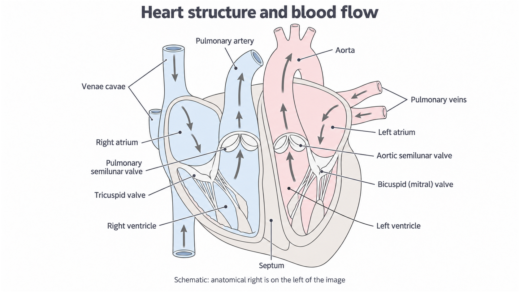
The anatomical right side appears on the left of this schematic. Arrows trace the two separate blood routes; the drawing represents connections rather than a measured specimen.
The atrial walls are relatively thin because atria move blood a short distance into the nearby ventricles at low pressure. Both ventricles have thicker muscular walls because they must generate enough pressure to drive blood through a circuit. The left ventricular wall is thicker than the right ventricular wall: the systemic circuit offers greater resistance to flow, so the left ventricle must generate higher pressure. The right ventricle pumps through the lower-resistance pulmonary circuit at lower pressure, helping protect the delicate lung capillaries.
The two ventricles normally pump the same volume per beat. The thicker left wall produces a greater pressure, rather than continuously sending more blood than the right side. Unequal sustained outputs would cause blood to accumulate in one circuit. Coronary arteries branch from the aorta and supply the cardiac muscle itself with oxygen and nutrients for respiration; blood inside the chambers is not the muscle's main oxygen supply.
Pressure-operated valves
Blood flows from higher pressure to lower pressure when an open pathway connects the regions. Contraction of cardiac muscle raises the pressure of blood inside a chamber; relaxation lowers it. A valve responds to the pressure difference across it, making an available pathway one-way.
The atrioventricular valves lie between atria and ventricles. The right valve is the tricuspid valve; the left is the bicuspid valve, also called the mitral valve. They open when atrial pressure is greater than ventricular pressure. As ventricular pressure rises above atrial pressure, blood pushes their flaps together, closing the route back into the atria.
The flaps are connected by tough tendinous cords, or chordae tendineae, to papillary muscles in the ventricular wall. During ventricular contraction these supports stop the closed flaps turning inside out into the atria. They do not pull the valves open and they do not act as a second pump.
The semilunar valves guard the exits of the ventricles: the pulmonary valve is at the entrance to the pulmonary artery, and the aortic valve is at the entrance to the aorta. They open when ventricular pressure exceeds pressure in the corresponding artery. When arterial pressure becomes higher, a small tendency for blood to flow backwards fills the pocket-shaped flaps and closes them, preventing continuing backflow into the ventricles. These valves have no tendinous cords.
This means that “ventricle contracting” is not enough to decide whether blood has left the heart. At the start of ventricular contraction, ventricular pressure may already exceed atrial pressure but still be below arterial pressure. Both sets of valves are then closed briefly. Blood leaves only after ventricular pressure exceeds arterial pressure.
Use the pressures on the two sides of a valve to decide its state. The heart muscle generates pressure; valves direct the flow that pressure can produce.
The same rule works on both sides of the heart, although the right side operates at lower pressures.
Sequence of the cardiac cycle
One heartbeat is a repeating sequence of contraction, ejection, relaxation and filling. Its stages overlap between chambers: while the ventricles contract, the atria can relax and receive returning blood.
Cardiac cycle
The sequence of events during one heartbeat. Systole means contraction of a chamber's muscle; diastole means relaxation.
Start with both atria and ventricles relaxed. Blood returns through the venae cavae and pulmonary veins. Atrial pressure is slightly higher than ventricular pressure, so the atrioventricular valves are open and blood enters the ventricles passively. Semilunar valves remain closed because arterial pressure is higher than ventricular pressure. Much of ventricular filling has therefore occurred before the atria contract.
Atrial systole
The atria contract together while the ventricles remain relaxed. Atrial pressure rises, moving additional blood through the open atrioventricular valves into the ventricles. The semilunar valves remain closed. Atrial systole completes ventricular filling; it does not move blood directly into the aorta or pulmonary artery.
Ventricular systole
The atria relax and the ventricles contract together. Rising ventricular pressure first exceeds atrial pressure and closes the atrioventricular valves. At this early stage the semilunar valves are also closed, so pressure rises without blood leaving the ventricles: this is isovolumetric contraction, meaning contraction at approximately constant ventricular volume.
As ventricular pressure rises above arterial pressure, the semilunar valves open. The right ventricle ejects blood into the pulmonary artery and the left ventricle ejects blood into the aorta. Atrioventricular valves remain closed because ventricular pressure is still above atrial pressure. The atria receive blood from veins during their relaxation. The ventricles do not become completely empty during ejection.
Cardiac diastole
The ventricles now relax, while the atria are also relaxed. Ventricular pressure falls below arterial pressure, closing the semilunar valves. Initially ventricular pressure is still above atrial pressure, so both valve groups are closed again: isovolumetric relaxation. As ventricular pressure falls further, below atrial pressure, the atrioventricular valves open and passive filling resumes. The next atrial systole then completes filling again.
| Stage | What produces the pressure change? | Consequence for flow |
|---|---|---|
| Atrial systole | Atria contract; ventricles remain relaxed | Additional blood enters ventricles through open atrioventricular valves |
| Ventricular systole | Ventricles contract; atria relax | Atrioventricular valves close first; semilunar valves open once ventricular pressure exceeds arterial pressure |
| Cardiac diastole | Ventricles relax; atria are also relaxed | Semilunar valves close first; atrioventricular valves open once ventricular pressure falls below atrial pressure |
Valves are therefore not fixed in one state for every moment of a named stage. The brief intervals with both groups closed explain how the ventricle changes its pressure before ejection or filling. The first main heart sound is associated with atrioventricular valve closure; the second is associated with semilunar valve closure.
Reading cardiac pressure traces
A pressure–time graph makes the sequence visible by showing when one pressure overtakes another. The graph below is an original simplified left-heart teaching model, not a patient recording. Its values and stage durations are illustrative; a real trace has additional small fluctuations and changes with heart rate and physiological conditions.
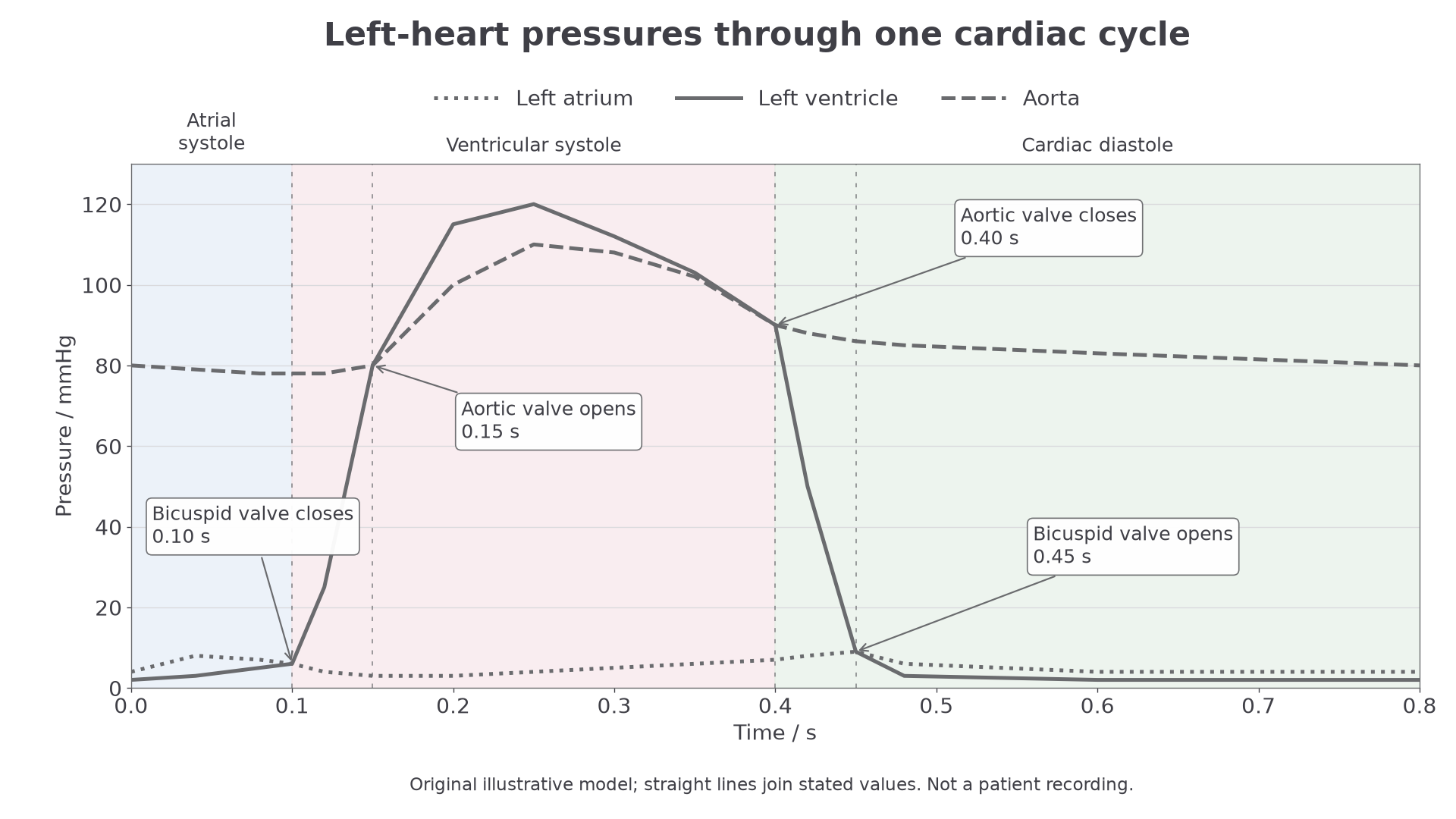
Read the vertical separation of the curves to identify a pressure gradient. The dashed event lines mark pressure crossings that change a valve's state; only the left-heart pressures are shown.
First identify the axes: time is in seconds and pressure in mmHg. Then compare the left ventricular curve with the left atrial curve for the bicuspid valve, and with the aortic curve for the aortic semilunar valve. A high aortic pressure alone does not mean blood is being ejected: ventricular pressure must be still higher.
For example, at 0.12 s this model has ventricular pressure 25 mmHg, atrial pressure 4 mmHg and aortic pressure 78 mmHg. The bicuspid valve is closed because 25 is greater than 4. The aortic valve is also closed because 25 is less than 78. This is the early closed-valve interval of ventricular systole. At 0.25 s the ventricular pressure is 120 mmHg and aortic pressure is 110 mmHg, so the aortic valve is open and ejection occurs.
The duration of an interval can be obtained from its two positions on the time axis.
Use the same time units for both readings.
In this model ventricular systole extends from 0.10 s to 0.40 s, giving . Ejection occupies only the interval from about 0.15 s to 0.40 s; it begins after the initial pressure rise. During diastole the aorta stays at a higher pressure than the ventricle because arterial elastic recoil maintains pressure as blood continues into the systemic circuit. Closing the aortic valve prevents that recoil from driving blood back into the heart.
At an exact crossing, pressures are approximately equal and a valve is changing state. Decide whether a valve is open or closed from the neighbouring interval, rather than claiming substantial flow from an equal-pressure instant.
Investigating heart structure
Observation and dissection can test whether features of a mammalian heart support the proposed pumping mechanism. A useful investigation asks whether the left ventricular free wall is thicker than the right, and examines how valves and their supports could maintain one-way flow. A free wall is the outer chamber wall, rather than the septum shared by the ventricles.
Preparation and observation
This is supervised laboratory work using a fresh heart supplied through an approved butcher or educational supplier, ideally with major vessels and atria intact. The teacher or technician completes the centre's risk assessment and demonstrates tool use. The apparatus includes a dissection tray, dissecting scissors, forceps, a blunt probe, a washable millimetre ruler and, where needed, a scalpel used under the teacher's supervision.
Before cutting, orient the specimen using the apex, chamber walls and vessel connections; do not assume its right side is on your right. Observe coronary vessels on the outer surface. Follow attached vessels gently with a blunt probe to establish which chamber they enter or leave. Do not force a probe through a wall or valve. A trimmed specimen may lack recognisable vessels or atria: record this limitation rather than guessing a connection.
The teacher demonstrates how to open each ventricle along a free wall, away from the septum and valve attachments, using shallow controlled cuts. Open the chamber carefully and follow it towards its artery. Examine the atrioventricular flaps, tendinous cords and papillary muscles, and the semilunar pockets at the arterial exits. Gently moving a flap can reveal the constraint imposed by its cords; stretching or cutting the cords would destroy the feature being investigated. Note the thinner atrial walls where these remain intact.
A fair wall-thickness comparison
For a paired comparison, the factor compared is chamber side, left or right; the measured outcome is ventricular free-wall thickness in millimetres. Use the same heart for each pair, so species, overall heart size and specimen condition are shared. Measure at corresponding levels midway between base and apex, using a cut surface perpendicular to the wall. Exclude external fat, the septum and internal papillary projections. Keep the ruler perpendicular to the cut surface and avoid compressing the soft tissue.
Take readings at several predefined, corresponding free-wall positions on each side and record the individual values, not only a mean. Keep anatomical level, measurement technique, ruler resolution and specimen hydration or preservation condition consistent. A ruler marked every 1 mm cannot justify wall measurements quoted to 0.01 mm. An ideal scale reading might have about ±0.5 mm uncertainty, but an uneven cut, deformable tissue and uncertain boundaries can make the actual uncertainty larger.
Here is an original illustrative dataset, supplied for processing practice rather than recorded from a real dissection:
| Corresponding position | Left wall / mm | Right wall / mm |
|---|---|---|
| 1 | 12 | 4 |
| 2 | 13 | 4 |
| 3 | 12 | 5 |
For each side, add its three readings and divide by three. For the left wall, to one decimal place; for the right wall, . Those extra decimal places do not improve the ruler's precision: biologically the result is approximately 12 mm compared with 4 mm. The separation is much larger than the ideal ruler reading uncertainty, supporting the conclusion that this specimen's left ventricular free wall is thicker. This is consistent with generating higher systemic pressure; wall thickness alone does not directly measure contraction force or blood pressure.
Several positions in one heart are repeated measurements of that specimen, not several independent animals. To investigate how general the difference is, repeat the paired method on several hearts of the same species and comparable size, keeping specimen preparation consistent. Preserve individual heart identity and compare paired results; averaging all readings without their identities can hide between-heart variation. An oblique cut overestimates thickness, and damaged or compressed walls can distort it.
Record labelled observations and an unshaded biological drawing of what is actually visible, using clear single lines, labels and measured scale where available. Distinguish an observation such as “thin flaps attached to cords” from the inference “cords constrain inversion during contraction”. A dissected heart is not beating, so it cannot by itself show living pressure changes or prove every valve's behaviour during the cardiac cycle. Models or pressure recordings supply that complementary evidence.
Safe and ethical handling
Sharp tools can cause cuts: keep material supported in the tray, cut away from fingers and follow the demonstrated technique. Raw tissue may carry microorganisms: cover skin cuts, avoid touching the face, wear protective clothing and gloves as directed, wash hands after handling, and clean equipment and surfaces using the centre's approved procedure. Use eye protection where splashes or cutting create an eye risk. Store specimens separately from food and dispose of tissue and sharps through the centre's designated routes; report injuries immediately to the supervisor.
Using tissue obtained as a by-product of the food industry avoids commissioning an animal's death solely for this lesson. Religious, cultural or personal objections still deserve an alternative such as observation of a teacher demonstration, a model or a recorded dissection. Online study prepares you to interpret and plan this investigation; it does not certify hands-on competence or the practical endorsement. The centre must observe and authenticate the relevant practical skills.