1.12, 1.13 - Carbohydrates and glycosidic bonds
Glucose can supply a cell with fuel, but storing thousands of separate glucose molecules would disturb its water balance. In 1.12, you will relate the structures of sugars and storage carbohydrates to their roles in providing and storing energy. In 1.13, you will explain how glycosidic bonds join sugar units and how hydrolysis splits them.
Single sugars and alpha-glucose
A carbohydrate may be one sugar unit, two joined units, or a polymer containing many units.
Monosaccharide
A single sugar unit that cannot be hydrolysed into smaller sugars. Glucose, fructose and galactose are examples.
Glucose, fructose and galactose each have the molecular formula , but their atoms are arranged differently. A molecular formula tells you the numbers of atoms, not their arrangement. Do not identify a sugar from its formula alone, or assume that every monosaccharide has six carbons.
In water, glucose occurs mainly as rings. The form used to build the storage carbohydrates in this lesson is α-glucose. Its six-membered ring contains five carbon atoms and one oxygen atom; the sixth carbon is in a group attached outside the ring. Several hydroxyl groups, , interact with water by hydrogen bonding. This helps explain why glucose is soluble and can be carried in aqueous fluids to cells. It is a small molecule that does not first need glycosidic-bond digestion before use as a respiratory substrate.
Reading its structure accurately
In the conventional Haworth view of α-D-glucose, place the ring oxygen at the upper right and C1 at the right. Follow the ring from C1 through C2, C3, C4 and C5, then back through oxygen. C6 belongs to the group attached to C5, rather than to the ring. The exact positions are:
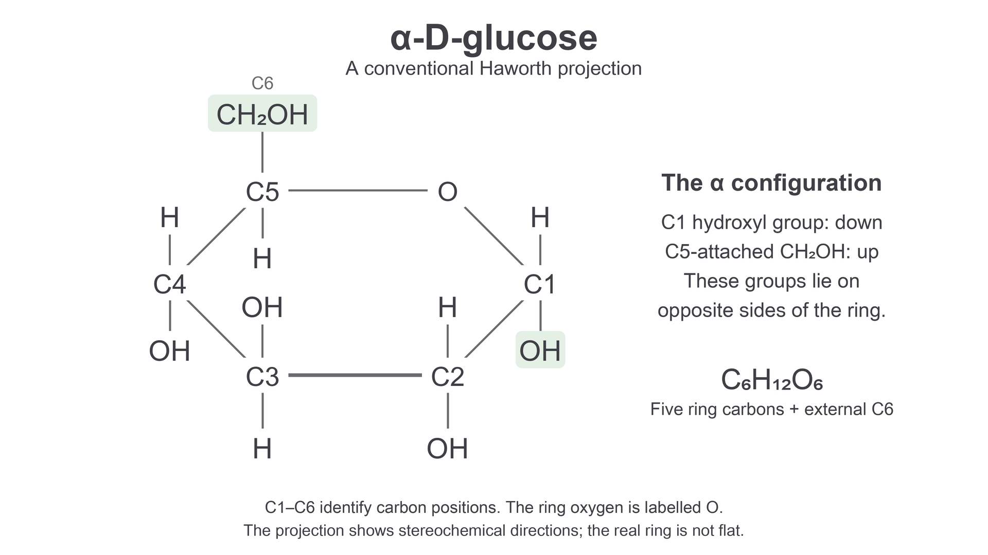
C1–C6 label carbon positions. In this conventional projection, the C1 hydroxyl group points down and the C5-attached CH₂OH group points up, on opposite sides of the ring. The external CH₂OH contains C6. The projection represents the molecule's stereochemistry rather than a flat physical ring.
| Position | Group and side of the ring in this view |
|---|---|
| C1 | Hydroxyl group below the ring |
| C2 | Hydroxyl group below the ring |
| C3 | Hydroxyl group above the ring |
| C4 | Hydroxyl group below the ring |
| C5 | group above the ring |
Thus, the C1 hydroxyl group and the group lie on opposite sides: this identifies the α form. Hydrogen occupies the opposite direction at each of C1–C5. A Haworth view simplifies the molecule's three-dimensional shape; the real ring is not flat. The β-glucose and cellulose structures belong to Topic 4, rather than this Topic 1 lesson. ChEBI's α-D-glucose record verifies the molecular identity.
Joining two sugar units
Two monosaccharides can join to make a larger molecule, but two separate sugars mixed together are still two separate molecules.
A disaccharide contains two monosaccharide units joined by a glycosidic bond, a covalent linkage through an oxygen atom. In the net condensation reaction, the joining groups lose the elements of one water molecule: an OH from one group and an H from the other form . The remaining oxygen links the sugar units. One new glycosidic bond corresponds to one water molecule formed in this chemical summary.
For a disaccharide formed from two sugars with formula :
The atom count balances: the products together contain 12 carbons, 24 hydrogens and 12 oxygens. The disaccharide alone contains fewer hydrogen and oxygen atoms than the two free sugars, because some are now in water.
The joining sugars and the positions of the bond determine which disaccharide forms:
| Disaccharide | Joining monosaccharides | Exact linkage |
|---|---|---|
| Maltose | Glucose + glucose | α(1→4): C1 of the first glucose links through oxygen to C4 of the second |
| Lactose | Galactose + glucose | β(1→4): C1 of galactose links through oxygen to C4 of glucose |
| Sucrose | Glucose + fructose | C1 of α-glucose links through oxygen to C2 of fructose; the linkage is α(1↔2)β |
The numbers specify the linked carbon positions; the α or β label specifies the configuration at the sugar's joining carbon. The β label in lactose refers to the galactose joining carbon, not a requirement to study β-glucose here. All three named disaccharides have formula , so their common formula does not make them the same molecule. These exact linkages are checked against ChEBI's maltose, lactose and sucrose structures.
Disaccharides are soluble sugars. Sucrose is used to transport carbohydrate in plants, and lactose supplies carbohydrate in milk. In human digestion, their glycosidic bonds must be split into monosaccharides before absorption. Those sugars can then enter metabolic pathways that provide energy. Dissolving a disaccharide in water is not the same as splitting its bond.
Splitting bonds with water
Hydrolysis reverses the net joining reaction by using water to split a glycosidic bond.
Hydrolysis
Splitting a chemical bond by reaction with water. For each glycosidic bond hydrolysed, one water molecule is consumed and its H and OH restore groups on the separated sugar units.
For example, complete hydrolysis of maltose uses one water molecule and gives two glucose molecules. Lactose gives glucose and galactose; sucrose gives glucose and fructose. Suitable enzymes catalyse these reactions in digestion: maltase acts on maltose, lactase on lactose and sucrase on sucrose. The products depend on the starting structure, rather than on the enzyme creating new kinds of sugar.
This formula equation fits each named disaccharide, but the sugar names are needed to distinguish the products. Complete hydrolysis splits all the relevant bonds; partial hydrolysis of a long chain can leave shorter chains or disaccharides. Neither dissolving nor hydrolysis alone is cellular respiration. Hydrolysis makes smaller substrates available; subsequent respiration transfers energy from fuel molecules to ATP through a series of reactions. Breaking a bond by itself requires energy, so “the bond breaks and releases energy” is not a sufficient explanation.
Counting joining and splitting events
Start with four separate glucose molecules. Joining the first two makes one bond, adding the third makes a second, and adding the fourth makes a third. A single, open chain of four residues therefore has three glycosidic bonds and its formation is summarised by the loss of three water molecules. Residue means the part of a sugar molecule incorporated into the joined carbohydrate. Complete hydrolysis of that chain consumes three waters and restores four glucose molecules.
More generally, one connected carbohydrate made from monosaccharides, with no closed loops, has joining bonds. Branching changes where those bonds are, but not this count. For example, an open branched molecule containing six residues still has five joining bonds. This rule describes individual molecules; it must not be applied unchanged to several disconnected chains or a closed ring of residues. The condensation equation is a net chemical model; cellular polymer synthesis uses enzyme pathways and activated sugar donors.
Amylose, amylopectin and glycogen
Repeating glycosidic linkages joins many sugar residues into a polysaccharide, and the linkage positions determine whether it branches.
A polysaccharide is a carbohydrate polymer containing many monosaccharide residues. Starch is the main carbohydrate reserve in plants and contains two kinds of glucose polymer, amylose and amylopectin. These are different molecules within starch, rather than alternating names for one chain. Glycogen is an important glucose reserve in animals, especially in liver and skeletal muscle.
| Polymer | Glucose linkages | Chain structure |
|---|---|---|
| Amylose | α(1→4) | Unbranched chain that can coil into a helix |
| Amylopectin | α(1→4) along chains; α(1→6) at branch connections | Branched chains |
| Glycogen | α(1→4) along chains; α(1→6) at branch connections | More frequent branching than amylopectin |
In an α(1→4) linkage, C1 of one glucose residue links through oxygen to C4 of the next. In an α(1→6) branch linkage, C1 of a residue on the new branch links to C6 of a residue in the existing chain. The branch connection is still a glycosidic bond. It is not a hydrogen bond or an extra glucose unit floating beside the chain. ChEBI verifies amylose's unbranched α(1→4) structure and amylopectin's chain and branch linkages.
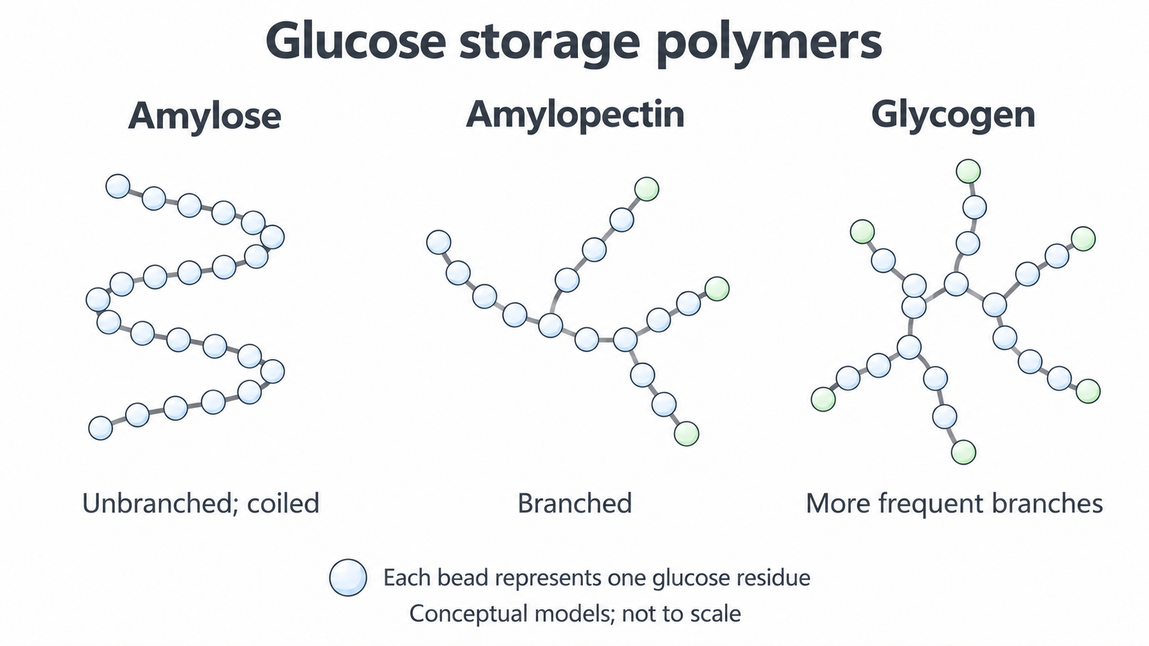
Each bead represents a glucose residue and each connecting line a glycosidic linkage. These simplified models show connectivity and relative branching, not exact residue counts, branch spacing or molecular dimensions. The table specifies the exact bond types.
All three polymers can yield glucose on complete hydrolysis. Thus, finding only glucose after hydrolysis does not identify which storage polymer was present: evidence about branching and linkages is also needed. Conversely, a branched glucose polymer cannot be amylose in the unbranched model used here. The structures provide a reusable reserve of sugar units, rather than a new kind of glucose that cannot be used for respiration.
Why polymer structure suits energy storage
A useful carbohydrate reserve must hold many glucose units inside a cell and make them available when demand increases.
Free glucose is useful for immediate supply because it is small, soluble and available as a respiratory substrate. These same properties create a storage problem. Thousands of separately dissolved glucose molecules produce many solute particles, encouraging water to enter by osmosis and potentially disturbing cell volume. Osmosis is the net movement of water through a partially permeable membrane from a more dilute to a more concentrated solution of solutes.
Linking glucose into large polymers stores many units in far fewer independently moving particles. Starch is stored in insoluble granules, while glycogen forms large hydrated particles. They are retained inside cells and contribute much less to osmotic effects than the equivalent number of free glucose molecules. Avoid saying that a polymer has absolutely no interaction with water or no osmotic effect; the important comparison is with the same glucose reserve stored as separate soluble sugars.
Shape also matters. Amylose's coiling packs a long chain into a smaller space. Amylopectin and glycogen's branched arrangements provide compact reserves and many chain ends. Several enzymes can act at different accessible ends at the same time, allowing rapid mobilisation of sugar units. Glycogen's more frequent branching gives more such ends for a comparable number of glucose residues, suiting the changing energy demands of animal cells. The speed of mobilisation also depends on the enzymes and conditions, so branching alone does not give a universal rate for every sample.
Muscle glycogen supplies fuel for that muscle's respiration. Liver glycogen contributes to maintaining a supply of glucose in blood between meals. Plant starch can provide glucose substrates for respiration during growth or when photosynthesis is unavailable. In every case, the stored carbohydrate becomes useful through controlled enzyme reactions and respiration. Glycosidic bonds can be hydrolysed, but animal glycogen mobilisation also uses phosphate-dependent cleavage reactions; it is not entirely direct hydrolysis to free glucose. Detailed pathway chemistry is not needed to explain the storage adaptation. The branch-end role and tissue distinction are supported by NCBI's glycogen account.
Storage suitability follows a chain of reasons: polymerisation reduces the number of separate solute particles, compact shape holds many units in little space, and branching supplies many accessible ends for controlled mobilisation. Respiration of the resulting substrates, rather than bond breaking alone, provides usable energy.
This explains why a cell can keep a substantial reserve without maintaining the same amount as freely dissolved glucose. It also explains why “large” and “branched” are useful only when their consequences are stated.
Inferring carbohydrate structure from evidence
Use product identity and linkage evidence together, because one observation may fit several carbohydrates.
The following is an original NovaLearn practice dataset, constructed to compare the models in this lesson; it is not a report of measured natural samples.
| Sample | Products of complete hydrolysis | Linkages present before hydrolysis | Branching evidence |
|---|---|---|---|
| P | Glucose only | α(1→4) only | Unbranched |
| Q | Glucose only | α(1→4) and α(1→6) | Branched |
| R | Glucose only | α(1→4) and α(1→6) | More branch connections than Q for the same number of residues |
P is consistent with amylose because it combines glucose-only products with an unbranched α(1→4) chain. The glucose-only result alone would also fit amylopectin or glycogen, so it is insufficient evidence on its own. Q and R share the same linkage types; R's greater branching is consistent with the glycogen model compared with amylopectin. To compare polymers, give both a shared feature and a difference, rather than describing only one sample.
The evidence predicts more accessible chain ends in R for the same glucose reserve. Under otherwise comparable conditions, that can support faster mobilisation by enzymes acting at those ends. The table does not measure rates, specify enzyme amounts or prove how quickly a real food is digested. A justified inference states both the structural reason and the conditions under which the prediction applies.