3.1, 3.2 - Cells and eukaryotic ultrastructure
In 3.1, you will connect the common features of cells to the needs of living organisms. In 3.2, you will learn how the internal structures of eukaryotic cells carry out different jobs, and use those structures to make reasoned predictions about a cell's activity.
The shared cellular plan
All living organisms consist of one or more cells: a bacterium is a single cell, while an animal contains many interacting cells.
Cell
The basic structural and functional unit of a living organism.
Cells share a common organisational plan. A cell-surface membrane separates the cell's contents from its surroundings and controls exchange. Cytoplasm contains the cell's internal structures and the aqueous material in which many chemical reactions occur. DNA stores inherited information, including instructions for making proteins, and ribosomes make those proteins. A cell needs both information and a way to use it; DNA alone cannot carry out the reactions encoded by its genes.
This is a common plan across cellular life, rather than a claim that every specialised mature cell retains every structure. For example, a mammalian red blood cell loses its nucleus and ribosomes during development; it was produced from a precursor cell containing them. Viruses are acellular particles, rather than organisms built from cells.
A eukaryotic cell normally has its main DNA enclosed in a nucleus. Animals, plants, fungi and protoctists are eukaryotes. Membranes also divide a eukaryotic cell into internal compartments. Each compartment can contain a different mixture of enzymes and maintain conditions suited to its reactions. The cytosol is the fluid part of the cytoplasm; in a eukaryote, cytoplasm refers to the contents outside the nucleus.
Organelle
A specialised structure within a cell that performs a particular function. Some organelles are membrane bound; ribosomes and centrioles are not.
Ultrastructure means the fine detail of cell structure, much of which requires electron microscopy to resolve. The diagram is an enlarged conceptual map of these structures, not an electron micrograph or a source of measurements.
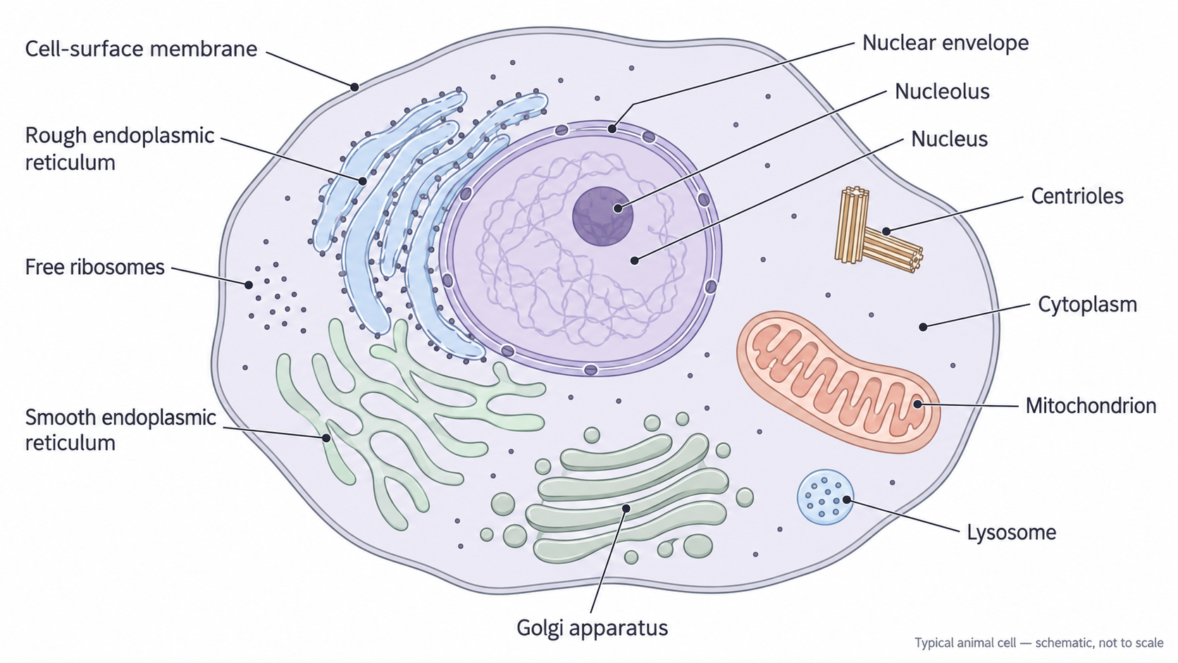
A typical animal cell, drawn schematically and not to scale. Organelle number, size and position vary between cells; plant cells also have many of these organelles.
The nucleus, nucleolus and ribosomes
The nucleus houses the main genetic information, while ribosomes use genetic instructions to assemble proteins.
The nucleus is surrounded by a nuclear envelope, made of two membranes. Nuclear pores provide controlled routes between the nucleus and cytoplasm: messenger RNA can leave, while proteins needed in the nucleus can enter. Inside, DNA associated with proteins forms chromatin; it becomes more condensed into visible chromosomes during cell division. Genes are expressed to make RNA and proteins, so nuclear DNA influences cell structure and activity through the proteins produced. Saying that the nucleus “controls the cell” is shorthand for these effects, rather than a separate controlling force.
The nucleolus is a dense region inside the nucleus, without its own surrounding membrane. Ribosomal RNA is made there and combined with proteins to assemble ribosomal subunits. These subunits leave through nuclear pores and join to form functioning ribosomes. The nucleolus makes ribosomal components; it is not the site where all the cell's proteins are made.
A ribosome is made of ribosomal RNA and protein, arranged into a large and a small subunit. It has no membrane. During translation, it reads messenger RNA and helps join amino acids into a polypeptide. Free ribosomes in the cytosol make many proteins used within the cell, including cytosolic enzymes. Other ribosomes become attached to the rough endoplasmic reticulum while making proteins that enter that membrane system. These are locations of the same kind of cytoplasmic ribosome, rather than permanently different types.
Endoplasmic reticulum and Golgi apparatus
The endoplasmic reticulum and Golgi apparatus provide membrane surfaces and enclosed spaces for making and processing cell products.
The endoplasmic reticulum (ER) is an interconnected membrane system enclosing an internal space called the lumen. Its membrane is continuous with the outer membrane of the nuclear envelope. Rough ER (rER) usually includes flattened membrane sacs called cisternae, with ribosomes attached to the cytosolic face. The attached ribosomes produce polypeptides that enter the ER lumen or membrane; proteins in the lumen can fold and begin processing. Its extensive membrane provides space for many attached ribosomes and associated proteins. Rough ER does not itself read messenger RNA: the ribosomes do that.
Smooth ER (sER) is a more tubular membrane network without attached ribosomes. Its enzymes synthesise lipids, including phospholipids; specialised cells also use it in steroid production or detoxification. Smooth ER can store calcium ions, especially in muscle cells. Its extensive membrane provides a large surface for these membrane-associated enzymes. “Smooth” describes the absence of attached ribosomes, rather than an absence of chemical activity.
The Golgi apparatus consists of a stack of curved, flattened membrane sacs, with small vesicles near its edges. A vesicle is a small membrane-bound compartment. The Golgi modifies, sorts and packages proteins and lipids for particular destinations. For example, adding or modifying carbohydrate groups helps produce glycoproteins. Its separate sacs contain different processing enzymes; vesicles allow selected products to be packaged without dispersing them through the cytosol. The Golgi has no attached ribosomes and does not synthesise polypeptide chains. The detailed route connecting rough ER, Golgi and secretion is developed in 3.3.
Ribosomes synthesise polypeptides; rough ER provides a compartment for entry, folding and initial processing; smooth ER has roles such as lipid synthesis; the Golgi modifies, sorts and packages products. A named “protein-making organelle” is not an adequate explanation of all four roles.
The visible roughness of ER and the smooth stack of Golgi sacs therefore reflect different activities, rather than interchangeable versions of the same organelle.
Continue this lesson.
You’re previewing NovaLearn without an account. Create a free account to read the rest.
Create free account