7.10 - Muscle fibres and performance
A skeletal muscle contains specialised cells that generate force, but these cells do not all contract at the same speed or resist fatigue equally well. Learn how a muscle fibre is organised, then connect its contractile proteins, membranes and ATP supply to the demands of sustained activity and rapid powerful movement.
From whole muscle to one cell
A whole skeletal muscle is an organ containing muscle tissue, connective tissue, blood vessels and nerves.
Within the muscle, connective tissue surrounds bundles called fascicles. Each fascicle contains many muscle fibres. Despite the word fibre, each muscle fibre is one long cylindrical cell, not a collection of separate cells lined up end to end. Connective tissue surrounds individual fibres as well as bundles, supports the tissue and transmits force towards the tendons.
Muscle fibre
A long, specialised skeletal muscle cell containing many nuclei and many parallel myofibrils.
During development, precursor cells fuse to form a muscle fibre. The mature fibre therefore has multiple nuclei, usually near its surface just beneath its cell membrane. These nuclei support gene expression and protein production across a large cell. Its cell membrane is called the sarcolemma, and its cytoplasm is the sarcoplasm.
The sarcoplasm contains many long contractile threads called myofibrils, running along the fibre. Each myofibril consists of a series of repeating units called sarcomeres. Keep the levels separate: a fibre is a cell; a myofibril is a structure inside that cell; a sarcomere is a repeating section of a myofibril.
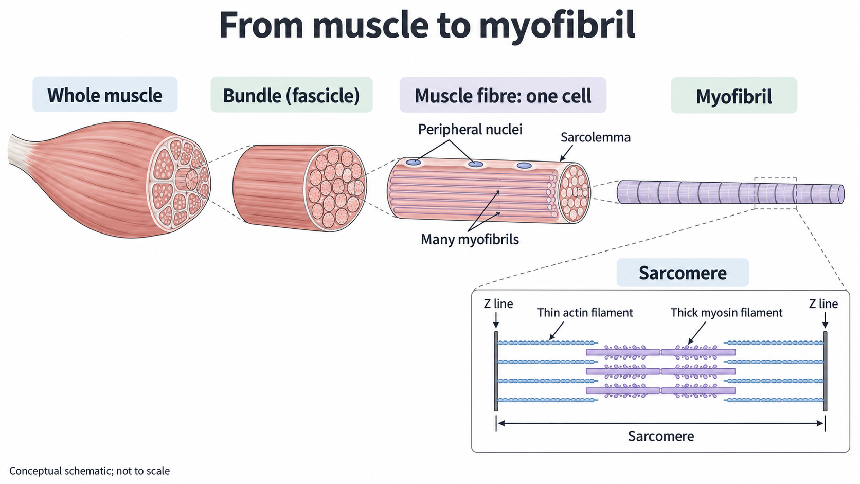
The enlargement sequence shows the nested organisation of skeletal muscle. The sarcomere inset introduces the thin actin and thick myosin filaments described later; the drawing is conceptual and not to scale.
Membranes and ATP supply
A muscle fibre needs to activate contractile structures throughout its thickness and continually replace the ATP it uses.
The sarcolemma folds inwards to form transverse tubules, usually shortened to T tubules. These carry electrical excitation from the surface deep into the fibre, close to the myofibrils. This arrangement helps different regions of a large cell respond together rather than only the surface region responding first.
A separate membrane network, the sarcoplasmic reticulum (SR), surrounds the myofibrils. It stores calcium ions and releases them into the sarcoplasm when the fibre is activated. Calcium ions enable contraction; their removal back into the SR allows relaxation. The T tubule is continuous with the sarcolemma, whereas the SR is a distinct intracellular compartment. They lie close together but their internal spaces are not one continuous tube.
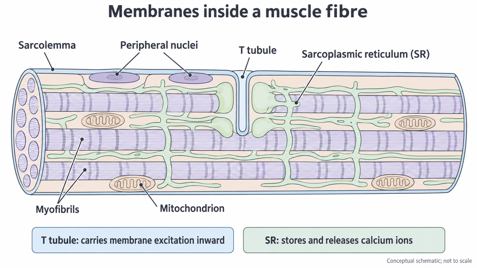
The T tubule carries membrane excitation inward; the separate SR stores and releases calcium ions. Mitochondria lie among the contractile structures. This is a schematic, not a micrograph or a measured cross-section.
Mitochondria provide sites for much of aerobic ATP production. ATP is also made by glycolysis in the sarcoplasm. Glycogen stored in the fibre supplies carbohydrate that can be broken down for respiration. ATP supports both contraction and active transport, including pumping calcium ions back into the SR. The energy available from ATP hydrolysis comes from the net reaction and its coupling to these processes; breaking a bond alone does not release energy.
Blood flows through capillaries outside the muscle fibres, delivering oxygen and respiratory substrates. Inside a fibre, myoglobin is an oxygen-binding protein. It provides a local oxygen reserve and helps oxygen reach sites of use; it is distinct from haemoglobin in red blood cells.
Sarcomeres and striations
The repeating arrangement of protein filaments within myofibrils gives skeletal muscle its striped appearance.
Sarcomere
The repeating contractile unit of a myofibril, extending from one Z line to the next Z line.
Each sarcomere contains thin filaments, mainly actin, and thick filaments, mainly myosin. Thin filaments attach to the Z lines at the ends and extend towards the middle. Thick filaments occupy the central region. Myosin has projecting heads that can interact with actin. The regular arrangement includes regions where the thin and thick filaments overlap.
The A band spans the length of the thick filaments and looks darker overall. It includes overlap regions. The I band contains thin filaments without thick filaments and is crossed by a Z line. The H zone, when visible, is the central part of the A band containing thick filaments without thin filaments. The M line lies at the centre, where proteins help hold the thick filaments in position. These are regions within the filament arrangement, not separate membranes.
Sarcomeres repeat along each myofibril, and the bands of neighbouring myofibrils align. This produces the transverse striations seen across a muscle fibre. A single myofibril is therefore much more organised than a loose mixture of actin and myosin molecules.
Force is generated through interactions between actin and myosin. As sarcomeres shorten, the filament overlap changes; the filaments themselves do not shorten. The calcium regulation and ATP-driven sequence of those interactions are developed in the sliding filament lesson.
Continue this lesson.
You’re previewing NovaLearn without an account. Create a free account to read the rest.
Create free account