8.1, 8.2 - Neurones, reflexes and pupil responses
How does a change outside the body make a muscle respond? In 8.1 you will connect the structures of sensory, relay and motor neurones to their jobs, including the role of Schwann cells and myelin. In 8.2(i–ii) you will follow nervous pathways from stimulus to effector, then explain how opposing iris muscles change the pupil in bright and dim light.
From stimulus to effector
A nervous response begins when a receptor detects a change and ends when an effector changes its activity.
Stimulus
A detectable change in the internal or external environment, such as a change in temperature, pressure or light intensity.
A receptor is a specialised cell or sensory nerve ending that detects a particular type of stimulus. Detection changes electrical signalling in a nervous pathway. A neurone is a specialised cell that transmits electrical signals called nerve impulses. A receptor is therefore the input to a pathway; it is not necessarily a whole sense organ. The eye, for example, contains many light-sensitive receptor cells.
In a mammal, sensory neurones carry information from receptors towards the central nervous system (CNS), which consists of the brain and spinal cord. A coordinator receives and processes information and organises an appropriate output. The CNS carries out this coordination. Motor neurones carry signals from the CNS to effectors: muscles that contract or glands that secrete substances. The changed activity of an effector produces the response.
The general sequence is:
Stimulus → receptor → sensory neurone → coordinator → motor neurone → effector → response.
These terms describe different roles. When a temperature receptor detects a rise in temperature and a sweat gland releases sweat, the gland is the effector. It performs the output; it does not detect the original temperature change or coordinate the whole pathway. Different nervous systems can organise pathways differently, but the distinction between detecting a stimulus, coordinating information and producing a response remains useful.
Three neurone specialisations
Sensory, relay and motor neurones share a basic cell organisation, but the arrangement of their processes suits different jobs in a pathway.
The cell body contains the nucleus and most of the organelles that maintain the cell and make its proteins. Branched dendrites receive input from receptors or other neurones, while an axon carries impulses towards its terminals. Terminal branches allow one neurone to communicate with other cells at junctions called synapses. A neurone is one cell; a nerve is a bundle of nerve fibres, mainly axons, supported by other tissues.
The common body-sensory neurone shown below has its cell body on a short side branch. The long process on the receptor side is conventionally called the dendron in school diagrams; the process leading to the CNS is called the axon. Together they carry the impulse from the receptor towards the CNS without the impulse needing to travel into and back out of the side-branch cell body. This is a typical arrangement, rather than the shape of every sensory neurone in every sense organ.
A relay neurone, also called an interneurone, lies within the CNS. Its branching inputs and usually relatively short axon allow connections between nearby neurones, including sensory and motor neurones in a simple reflex circuit. More complex CNS circuits contain many relay neurones.
A typical motor neurone has many short dendrites around a cell body in the CNS and a long axon extending to an effector. The dendrites allow it to receive inputs from other neurones. Its long axon carries the output over the distance from the CNS to a muscle or gland; terminal branches connect it with target cells. The neurone does not pull the muscle: its signal changes the muscle's activity.
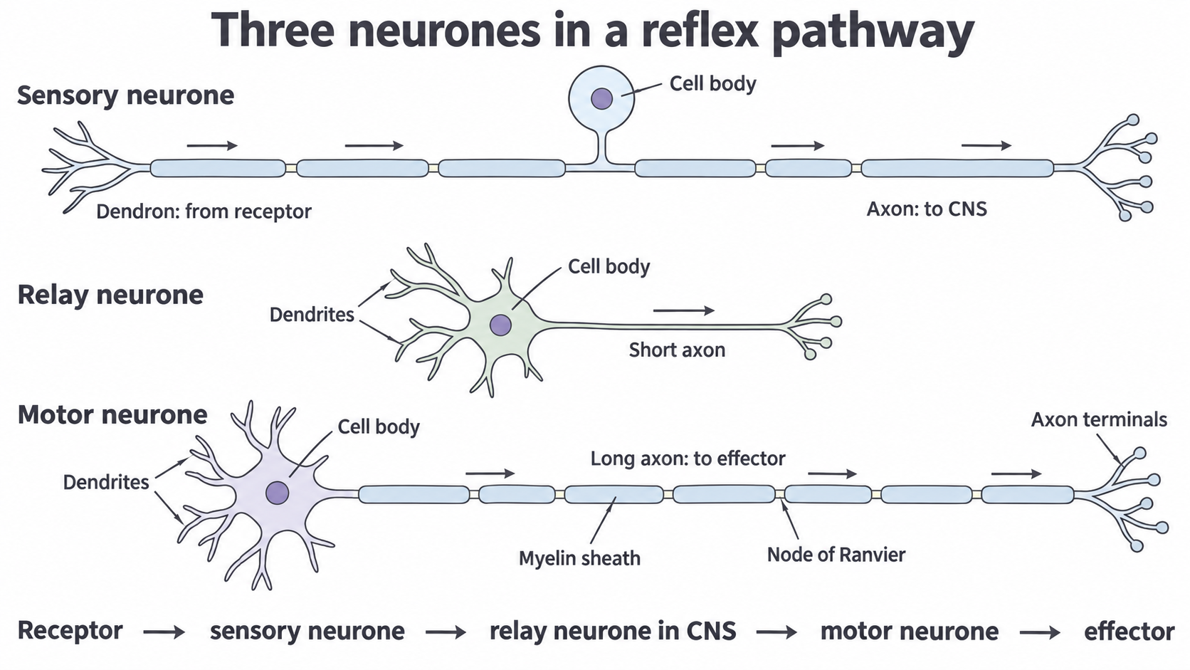
Typical neurone arrangements in a three-neurone reflex pathway. Arrows show impulse direction; the drawings are schematics and are not to scale.
Schwann cells and myelin
An impulse travelling along a long peripheral axon needs to reach its target quickly, and a myelin sheath helps it do so.
Schwann cells are supporting cells of the peripheral nervous system, outside the brain and spinal cord. A myelinating Schwann cell wraps its cell membrane repeatedly around one short length of an axon. The tightly packed, lipid-rich membrane layers form a segment of the myelin sheath. Many Schwann cells are needed to cover a long axon; one Schwann cell does not make the entire sheath along it.
Myelin acts as an electrical insulator, reducing loss of current across the covered axon membrane. Small gaps between successive sheath segments are nodes of Ranvier, where axon membrane is exposed. The combination of insulating segments and exposed nodes allows impulses to be conducted more rapidly than along an otherwise comparable unmyelinated axon. The detailed ion movements that generate and propagate an action potential are studied in 8.3; here the important structure–function link is insulating sheath → faster transmission over a long distance.
Myelin is not the axon's cytoplasm, and a Schwann cell is not another neurone. Not all axons are myelinated. Also, Schwann cells make peripheral myelin: the CNS has a different type of myelin-forming supporting cell. This prevents the mistaken inference that every myelinated process inside the brain is wrapped by Schwann cells.
The axon carries the impulse. Schwann cells form insulating myelin segments around peripheral axons, with nodes between segments; myelination increases conduction speed.
Damage to the sheath can therefore delay or disrupt information transfer even if the receptor still detects a stimulus. It does not change a sensory neurone into a motor neurone: those names refer to their roles and connections.
Reflex coordination
A reflex is an automatic response to a stimulus, so it can begin without a conscious decision.
Continue this lesson.
You’re previewing NovaLearn without an account. Create a free account to read the rest.
Create free account