2.6 - Transcription and translation
A DNA base sequence can determine an amino acid sequence even though DNA and amino acids do not bind directly to one another. This lesson explains the two stages that connect them, and how to follow the information accurately from a DNA template to mRNA and then to a polypeptide.
Two stages in a eukaryotic cell
Protein synthesis uses an RNA copy of genetic information and then reads that copy to assemble amino acids in a particular order.
Transcription and translation
Transcription produces an RNA molecule using a DNA strand as a template. Translation uses the sequence of codons on mRNA to determine the sequence of amino acids assembled into a polypeptide.
Recall that a protein-coding gene contains the information for an amino acid sequence. A polypeptide is a chain of amino acids joined by peptide bonds. The detailed chemistry of amino acids and protein folding follows in the next lessons; here the question is how the cell chooses their order.
In a eukaryotic cell, such as a human cell, transcription of a nuclear gene happens in the nucleus. The resulting messenger RNA (mRNA) carries the information to a ribosome in the cytoplasm, where translation occurs. Ribosomes can be free in the cytoplasm or attached to rough endoplasmic reticulum. DNA remains in the nucleus: neither the gene nor one of its DNA strands travels to the ribosome.
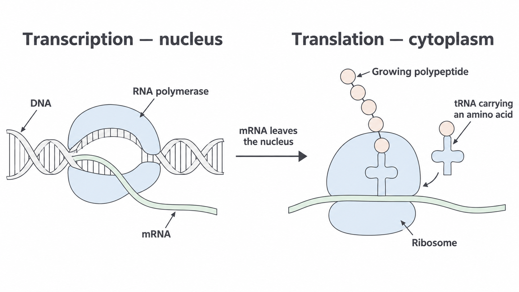
The two stages use different machinery. RNA polymerase makes an RNA copy; a ribosome uses that message while tRNA brings amino acids. This is a conceptual schematic, not a scale drawing or an exact base-pair diagram.
The information therefore passes DNA → mRNA → polypeptide. This arrow describes information transfer, not conversion of a DNA molecule into RNA or conversion of RNA into amino acids. The nucleic acids provide instructions; amino acids are separate building materials.
Transcription from the template strand
RNA polymerase assembles complementary RNA nucleotides in the order specified by one DNA strand.
The DNA template strand, also called the antisense strand, is the strand read during transcription of that gene. The opposite DNA strand is the coding strand, or sense strand. For a particular transcribed region, the coding strand has the same base order as the RNA when both are written in the same direction, except that DNA has thymine (T) where RNA has uracil (U). It is the template strand that is read; the name “coding strand” does not mean that RNA polymerase copies that strand.
The labels 5′ and 3′ distinguish the two ends of a nucleic acid strand. Complementary paired strands run in opposite directions: they are antiparallel. RNA polymerase reads the DNA template 3′ → 5′ and extends the RNA 5′ → 3′, adding each new RNA nucleotide to its 3′ end.
Building the RNA copy
- RNA polymerase associates with the DNA at the start of the region to be transcribed. A short section of the double helix opens: hydrogen bonds between complementary DNA bases are disrupted, exposing the template. The DNA sugar–phosphate backbones remain intact.
- Free RNA nucleotides pair with exposed template bases: DNA A pairs with RNA U, DNA T with RNA A, DNA C with RNA G, and DNA G with RNA C.
- RNA polymerase catalyses the joining of adjacent RNA nucleotides by phosphodiester bonds, forming the RNA sugar–phosphate backbone. Complementary pairing chooses the base order; backbone bonds make one continuous RNA strand.
- As polymerase moves along, the RNA separates from the template and the two DNA strands pair again behind it. At the end of transcription, the RNA transcript is released. After any necessary RNA processing, mRNA leaves the nucleus through a nuclear pore.
The following short fragment illustrates pairing, not a complete start-to-stop coding sequence. Each row is explicitly labelled and both ends are shown.
| Molecule | Sequence, left to right |
|---|---|
| DNA template strand | 3′–GAT CCA TGT–5′ |
| DNA coding strand | 5′–CTA GGT ACA–3′ |
| RNA transcribed from this template | 5′–CUA GGU ACA–3′ |
For example, the template triplet GAT gives RNA CUA by G–C, A–U and T–A pairing. The RNA matches the coding strand with U replacing T. Hydrogen bonds involved in base pairing must not be confused with phosphodiester bonds joining nucleotides along a backbone.
Translation at a ribosome
A ribosome reads the mRNA message while transfer RNA links each amino-acid instruction to the appropriate building material.
Codon and anticodon
A codon is a triplet of bases on mRNA that specifies an amino acid or a start/stop instruction. An anticodon is a triplet of bases on tRNA that can pair with a complementary mRNA codon.
Transfer RNA (tRNA) is a folded RNA molecule with an anticodon region and an amino-acid attachment site at its 3′ end. Before a tRNA brings an amino acid to the ribosome, enzymes attach the appropriate amino acid to it. Different tRNAs therefore act as adaptors between mRNA base triplets and particular amino acids. The amino acid attaches to tRNA at a different site from the anticodon; an amino acid does not base-pair with mRNA.
At the ribosome, a tRNA with a suitable anticodon pairs with the exposed mRNA codon through complementary base pairing. The anticodon and codon are antiparallel. For a simple example, mRNA 5′–GCU–3′ pairs with a tRNA anticodon 3′–CGA–5′. GCU specifies alanine in the standard genetic code, so this tRNA brings alanine. Codon tables are read using the mRNA codon, not the DNA template or tRNA anticodon. These examples use straightforward complementary pairing; details of alternative pairing are not needed here.
Extending the polypeptide
The ribosome binds mRNA and holds the interacting tRNAs in the correct positions. During elongation, an incoming tRNA presents the next amino acid alongside the growing chain. The ribosome catalyses peptide-bond formation, linking that amino acid into the polypeptide. Its ribosomal RNA is central to this catalytic role; the ribosome is more than a surface on which amino acids happen to meet.
The ribosome advances by one codon along the mRNA in the 5′ → 3′ direction. The tRNA that has given up its amino acid leaves and can be loaded again. Another suitable tRNA pairs with the next codon, and the cycle repeats. This gives a connected chain in the order instructed by successive mRNA codons, rather than a random mixture of the amino acids available in the cytoplasm.
The molecules have different jobs: mRNA supplies the ordered message; tRNA carries amino acids and recognises codons; the ribosome positions the molecules and catalyses peptide bonds. RNA polymerase works in transcription and does not join amino acids.
Start and stop codons
Translation must begin at an appropriate position and release the chain at its end, so not every mRNA triplet has the same role.
The usual start codon is AUG in the standard code used in these examples. During initiation, the ribosome and an initiator tRNA carrying methionine assemble at the start codon. Its anticodon pairs with AUG, and methionine becomes the first amino acid of the newly synthesised chain. Reading then continues in successive triplets. AUG also specifies methionine when it occurs within a coding sequence; it is its position and recognition during initiation that give a start codon its start role.
The standard mRNA stop codons are UAA, UAG and UGA. A stop codon does not specify another amino acid. When an in-frame stop codon reaches the decoding position, a protein release factor recognises it rather than an amino-acid-carrying tRNA. The completed polypeptide is released, and the translation machinery separates. You should not invent a tRNA that delivers a “stop amino acid”.
These signals control translation. AUG is not the signal that makes RNA polymerase begin transcription, and an mRNA stop codon does not itself tell RNA polymerase to stop copying DNA. Transcription has its own start and termination signals on DNA. An mRNA can also contain regions before and after its translated sequence.
The start codon establishes where translation begins; each following in-frame amino-acid codon supplies one amino acid; the first in-frame stop codon ends translation without adding an amino acid.
The chain initially made may subsequently be processed and folded into a functional protein. That later stage does not change the distinction between an amino-acid codon and a translation stop signal.
Following a sequence from DNA to polypeptide
A reliable sequence deduction starts by identifying the molecule, the strand and its direction before choosing any amino acid.
Here is an original simplified coding region with no introns. Its first AUG is the specified translation start; untranslated regions and RNA-processing steps are omitted. It is deliberately much shorter than a typical protein-coding sequence. Spaces show the translation triplets, not breaks in the molecules.
| Molecule | Sequence, left to right |
|---|---|
| DNA template strand | 3′–TAC CGA AAA ACT–5′ |
| DNA coding strand | 5′–ATG GCT TTT TGA–3′ |
| mRNA | 5′–AUG GCU UUU UGA–3′ |
Use this supplied excerpt of the standard genetic code. All codons are mRNA read 5′ → 3′.
| mRNA codon | Meaning |
|---|---|
| AUG | Methionine; start in this example |
| GCU | Alanine |
| UUU | Phenylalanine |
| UGA | Stop |
First, complementary pairing with the template gives AUG GCU UUU UGA. Checking against the coding strand gives the same order with U instead of T. Next, the ribosome reads from the indicated AUG. The chain is methionine → alanine → phenylalanine, in the order in which amino acids are incorporated. There are three amino acids: UGA releases the chain and is not a fourth amino acid.
For the three amino-acid codons, the simple complementary tRNA anticodons, written antiparallel to the mRNA, are 3′–UAC–5′, 3′–CGA–5′ and 3′–AAA–5′ respectively. There is no amino-acid-carrying tRNA anticodon to enter for the stop signal in this standard example.
Direction matters even for one triplet. If a DNA template triplet is given as 5′–CAT–3′, its complementary RNA aligned underneath is 3′–GUA–5′. Reading that RNA in the required 5′ → 3′ direction gives 5′–AUG–3′, not GUA. Equally, the anticodon aligned with 5′–AUG–3′ is 3′–UAC–5′; written in its own 5′ → 3′ direction, that anticodon is 5′–CAU–3′. Always show which direction you mean.