Core practical 11 - The Hill reaction in isolated chloroplasts
A blue dye lets us follow electron transfer in chloroplasts without waiting for a plant to grow. You will learn how to prepare a chloroplast suspension, make controlled measurements and use graphs to judge the evidence. A separate whole-leaf example will show why zero net gas exchange can occur while photosynthesis and respiration continue.
What DCPIP detects
The Hill reaction investigates light-driven electron transfer using chloroplast material separated from leaves. Recall that chlorophyll in thylakoid membranes absorbs light, exciting electrons. Water supplies replacement electrons through photolysis; electron transfer in the light-dependent reactions normally contributes to the reduction of NADP.
DCPIP
DCPIP is an artificial electron acceptor. Its oxidised form is blue at the buffered pH used here; its reduced form is colourless.
Adding DCPIP to an active chloroplast suspension gives electrons an artificial destination. Under suitable illumination, the blue contribution to the mixture decreases as DCPIP accepts electrons and is reduced. The chlorophyll remains green, so the final mixture need not look like colourless water. Compare it with chloroplast suspension lacking DCPIP.
This is an assay of the light-dependent electron-transfer system, rather than a direct measurement of glucose production. A fall in DCPIP absorbance does not show how much carbon dioxide was fixed, whether carbohydrate accumulated, or how rapidly an intact leaf was photosynthesising. The suspension contains disrupted cells and may include chloroplast fragments with active thylakoids. A working electron-transfer system does not require every chloroplast to retain an intact outer envelope.
In an intact plant, reduced NADP and ATP connect the light-dependent reactions to carbon fixation. Here DCPIP diverts electrons, and the preparation does not reproduce the complete intact-leaf system. Changing carbon dioxide availability therefore does not provide a direct test of the DCPIP-reduction step. A colour change alone also needs controls: other reducing substances released from leaves, or a change in the dye unrelated to chloroplast activity, could otherwise mislead us.
Preparing active chloroplast material
Extraction must release chloroplasts while retaining as much useful activity as possible. Use fresh, healthy green leaves from one species and a similar developmental stage. Record their source; use cultivated material and only the amount required. Avoid unnecessary plant damage and do not collect protected plants.
The apparatus includes a balance, scissors, chilled mortar and pestle, muslin and funnel, suitable centrifuge tubes and centrifuge, pipettes with a filler, an ice bath and labelled storage tubes. The teacher supplies a suitable isolation medium: a buffered solution with an appropriate osmotic concentration and ions. For example, a sucrose-containing phosphate buffer near pH 7 with potassium chloride is used in established school protocols. Sucrose helps reduce osmotic damage during isolation; buffer resists pH changes that could alter protein activity. Plain distilled water is unsuitable as the sole extraction medium because its osmotic conditions can damage the preparation.
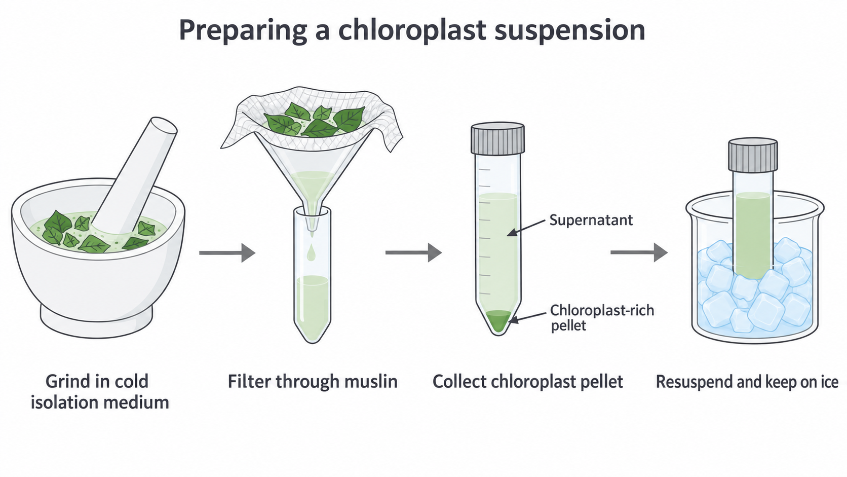
Preparation of a chloroplast-rich suspension. This is a conceptual illustration, not a microscope observation or a claim of complete purity.
Cut away tough stalks and midribs, weigh a recorded mass of leaf and grind it briefly in a measured volume of chilled isolation medium. Use the same mass-to-volume ratio for preparations being compared. Grinding breaks open cells; excessive or prolonged grinding can damage organelles and warm the mixture. Work promptly with chilled apparatus and keep the extract cold without freezing it.
Filter through several layers of pre-wetted muslin into a chilled vessel. The cloth retains large tissue debris while chloroplasts and smaller material pass into the filtrate. Filtration alone does not isolate pure chloroplasts. A suitable centrifugation step collects a chloroplast-rich pellet, leaving liquid called the supernatant above it. Follow the approved speed, time and rotor instructions for the actual centrifuge; an rpm value cannot be transferred between different rotor radii as though it guaranteed the same force. Balance opposed tubes by mass, secure the lid and wait for the rotor to stop completely before opening it.
Carefully remove the supernatant and gently resuspend the pellet in a measured small volume of cold medium. Keep this stock suspension on ice and use it promptly. Gently mix before removing each aliquot, because settling would otherwise give different chloroplast concentrations. A green supernatant may still contain chloroplasts or fragments; it is not automatically a no-chloroplast control.
Wear eye protection, use a pipette filler, handle blades and glass carefully, and clean dye splashes promptly. DCPIP stains and skin contact should be avoided. Use teacher-approved heating arrangements for any boiled control; cool it before comparison. Keep liquids away from lamp electrics, and handle hot lamps or water baths safely. A supervised local risk assessment and equipment instructions determine the actual procedure.
Comparisons and controls
A useful investigation changes one defined factor and measures how rapidly DCPIP loses absorbance. To investigate light intensity, select several measured intensities, including low and high values established by a pilot. Measure intensity at the reaction mixture with a suitable light sensor; lamp distance is only a way of adjusting light and is not itself a measurement of the light reaching the sample.
Keep wavelength, chloroplast concentration, DCPIP concentration, total volume, pH, optical path length and temperature constant. A water bath or suitable heat filter helps separate light effects from lamp heating; check the actual sample temperature. Allow reaction mixtures to reach the same chosen assay temperature before starting. The stock remains cold between trials, but all comparisons are measured at the same assay temperature. Begin timing at mixing, use the same mixing method and reading intervals, and keep any mixing-to-first-reading delay short and consistent.
The following is one cuvette-scale design. Volumes are illustrative and must fit the apparatus and a successful pilot. The DCPIP stock and its dye-free replacement use the same buffered solvent. Each mixture totals .
| Mixture | Chloroplast suspension / cm³ | DCPIP stock / cm³ | Isolation medium / cm³ | Dye-free stock solvent / cm³ | Treatment and purpose |
|---|---|---|---|---|---|
| Active light | 0.50 | 0.50 | 2.00 | 0 | Illuminated experimental reaction |
| Dark | 0.50 | 0.50 | 2.00 | 0 | Same active suspension, fully wrapped in foil; tests dependence on illumination |
| Boiled | 0.50 | 0.50 | 2.00 | 0 | Suspension heated sufficiently to disrupt activity, then cooled; tests dependence on heat-sensitive biological machinery |
| No chloroplasts | 0 | 0.50 | 2.50 | 0 | Medium replaces suspension; tests dye/medium change in light without the preparation |
| Optical blank | 0.50 | 0 | 2.00 | 0.50 | No dye; accounts for background from chloroplast material and other ingredients |
An experimental control tests an alternative explanation. The dark mixture asks whether illumination matters; the no-chloroplast mixture asks whether the dye changes in the medium alone. A boiled control supports the role of heat-sensitive structures or proteins, but boiling can also alter scattering or release compounds, so it is not proof of one named enzyme's involvement.
An optical blank serves a different purpose: it sets the colorimeter baseline for background absorbance and scattering without the measured dye. A blank is not an experimental negative control for light dependence. Match its composition to the relevant sample; a boiled or chloroplast-free mixture may need its own corresponding dye-free blank. Keep an illuminated dye-free preparation for a colour comparison and check that its background remains reasonably stable.
Use at least three fresh reaction mixtures at each intensity, rather than counting repeated readings of one cuvette as three independent trials. Randomise or alternate the order of intensities so that ageing of the stock is not confounded with light. The same stock limits preparation differences within a comparison; repeat with independent leaf preparations to assess wider biological repeatability. Keep dark samples protected during measurement: use an appropriate dark-compatible arrangement or separate protected samples for fixed exposure periods, because repeatedly unwrapping a tube illuminates it.
Measuring DCPIP reduction
Colorimetry replaces a subjective colour judgement with a quantitative reading. Select a wavelength or red filter at which oxidised DCPIP absorbs strongly, commonly near , using the instrument and pilot instructions. Blank the instrument with the matched dye-free mixture, and use clean matched cuvettes with the same path length, orientation and fill height. Wipe the clear faces, remove bubbles and avoid touching the optical surfaces. A fingerprint or settling particles can change a reading without any DCPIP reduction.
Use measured pipetted volumes, not estimated drops. Prepare a pilot to choose a chloroplast dilution and DCPIP concentration that yield a measurable change over the reading period while remaining in the colorimeter's usable range. For a twofold serial dilution, mix suspension with isolation medium; transfer of that dilution to another medium for a quarter-strength suspension. Mix each stage before transferring. Use one chosen dilution throughout the actual light comparison and record it; changing dilution between light treatments would change another factor.
Start the reaction by the final addition and mixing of DCPIP with chloroplast material. Record the start time and take absorbance readings at regular short intervals under the defined illumination. If the cuvette must be removed from the light to read it, standardise and minimise this interruption. A continuously recording arrangement must distinguish assay illumination from the measuring beam; the measuring beam itself can illuminate a nominally dark sample.
Absorbance is dimensionless, though instruments often display absorbance units or arbitrary units (AU). Within the calibrated working range, decreasing absorbance provides a proxy for decreasing oxidised DCPIP concentration. Converting the change to moles of DCPIP requires a suitable concentration calibration or extinction coefficient and path length; absorbance alone is not a molar rate.
Without a colorimeter, compare equal-depth tubes against a white background and record the time to reach one predefined colour standard. The chlorophyll-containing blank helps define loss of the blue component. An observer's endpoint is less precise than instrumental readings; use the same observer, lighting and endpoint rule. With the same initial amount of dye and the same endpoint in all tubes, in is a comparative rate index. It is not an initial gradient and does not have units of moles per second. A tube that fails to reach the endpoint within the observation period is reported as such, rather than assigned an invented zero rate.
Compare like with like: identical dye amounts and endpoints for timed decolourisation, or identical calibrated optical conditions and an appropriate time segment for absorbance rates.
Observing and drawing the preparation
A microscope checks what the preparation actually contains. Place a small drop of the well-mixed suspension on a clean slide and lower a coverslip gently to limit bubbles. Begin with low power, find the material and focus; then move to high power and use fine focus. Reduce the light if needed to improve contrast. Compare samples from the resuspended pellet and the supernatant using the same drop volume and viewing conditions. More green bodies in the pellet preparation supports enrichment, but does not establish purity or prove that those bodies are physiologically active.
Continue this lesson.
You’re previewing NovaLearn without an account. Create a free account to read the rest.
Create free account