3.10 - Mitosis and the cell cycle
A growing root and a healing skin wound both need new cells with a complete set of genetic instructions. This lesson explains how DNA copying, mitosis and cell division work together to produce daughter cells with the same chromosomes, and why this matters for growth and asexual reproduction.
Chromosomes and their copies
A cell must pass on a complete copy of its genetic information when it divides.
In a eukaryotic nucleus, DNA is organised into chromosomes, associated with proteins. Before DNA replication, each chromosome contains one long, double-stranded DNA molecule. Replication makes a second copy of that molecule. The resulting duplicated chromosome has two sister chromatids, each containing one DNA molecule. The sisters are held together, particularly at a region called the centromere, until they separate during mitosis.
Mitosis
The division of a eukaryotic nucleus in which duplicated chromosomes are separated to produce two daughter nuclei with the same chromosome complement as the original nucleus.
An X-shaped drawing represents one duplicated chromosome, with two sister chromatids; it does not represent two chromosomes. Nor is each arm of the X a separate chromatid: one entire side is one chromatid. The X shape is a compact form seen during division. Chromosomes still exist during interphase, even though their less condensed DNA is not usually visible as separate chromosomes with a light microscope.
Do not confuse sister chromatids with a homologous pair. The two homologous chromosomes carry the same genes at corresponding positions but may carry different alleles; one was inherited from each parent. Sister chromatids are copies of one of those chromosomes, so they normally carry the same alleles.
Ploidy describes the number of chromosome sets. A diploid nucleus, written , has two sets; a haploid nucleus, , has one. Copying each chromosome does not add a new set of different homologous chromosomes: a diploid cell remains diploid after replication. Its DNA quantity doubles because each chromosome now has two chromatids. Mitosis preserves the starting ploidy; in a diploid parent it produces diploid daughter nuclei.
Interphase prepares the cell
The cell cycle links the preparation of a cell to its division into two daughter cells.
Interphase is the period between successive nuclear divisions. It is active preparation, rather than a resting stage. Its three phases have different roles:
| Phase | What happens | Why it matters |
|---|---|---|
| G1 | The cell grows, synthesises proteins and increases its cellular components. | Division must be supported by enough cytoplasm and machinery for the daughter cells. |
| S | The nuclear DNA is replicated once. | Each chromosome acquires a sister chromatid, providing a copy for each daughter nucleus. |
| G2 | Further growth and preparation for division occur. | The cell prepares the proteins and structures needed to separate the copied chromosomes. |
DNA replication uses each original strand as a template for a complementary new strand. The result is two DNA molecules with the same base sequence, provided copying is accurate. This is why replication supplies matching sister chromatids, rather than a randomly assembled second set of instructions.
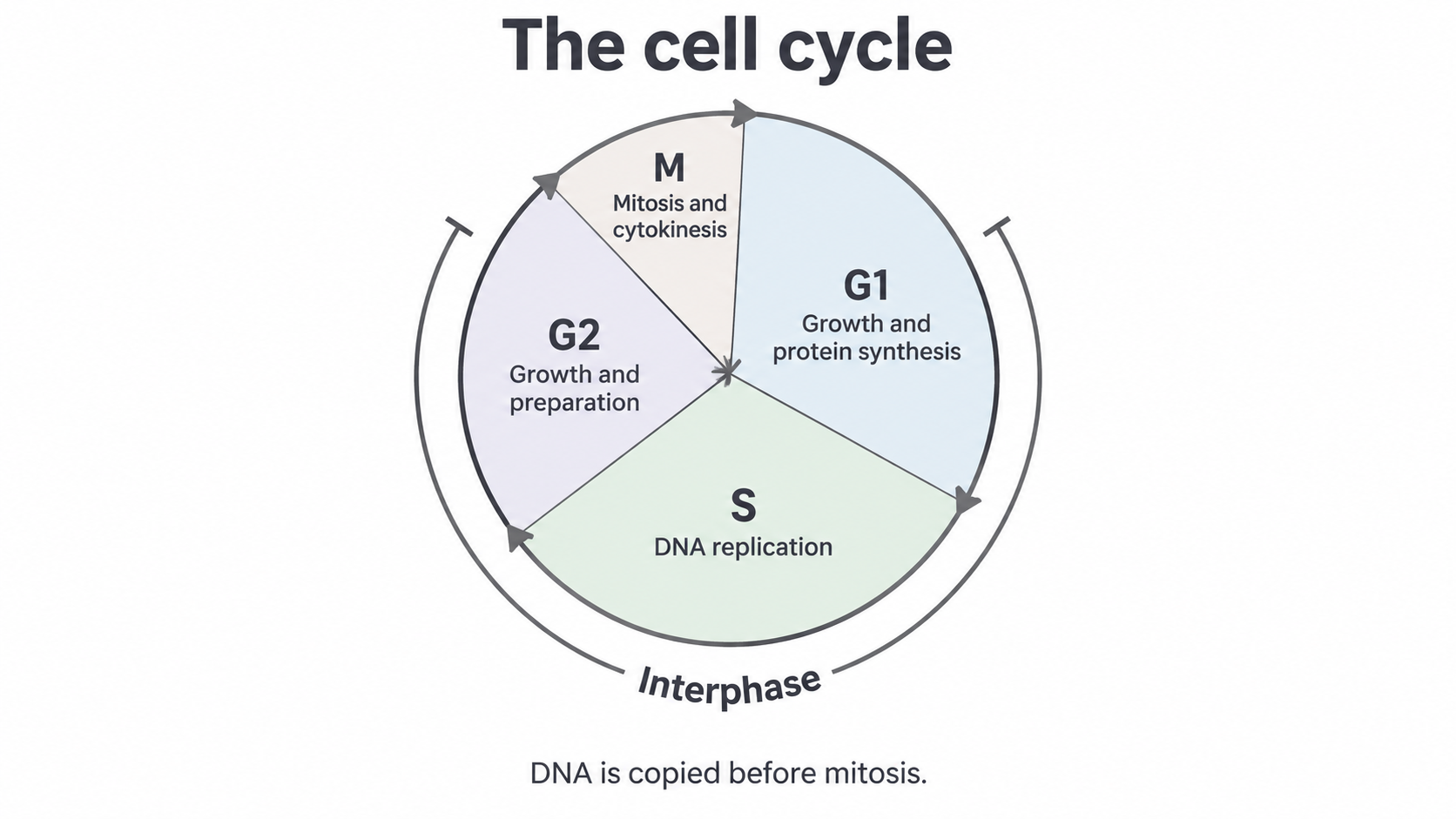
The cell cycle: G1, S and G2 make up interphase. DNA replication precedes mitosis. Sector sizes are schematic, not measured durations.
The M phase includes mitosis, which divides the nucleus, and cytokinesis, which divides the cytoplasm into separate cells. These events are linked but are different processes. Cytokinesis often starts before mitosis has completely finished. A daughter cell can enter G1 and repeat the cycle if it receives the appropriate conditions and signals. Many specialised cells do not keep dividing, so the diagram does not mean every cell cycles continuously.
The order is essential. Separating the chromosomes before copying the DNA would leave daughter cells without a complete chromosome complement. Copying DNA without then separating it would leave the extra copies in one cell. Growth between divisions also matters: repeated division without growth would make progressively smaller cells.
Separating the sister chromatids
Mitosis distributes one sister chromatid from every duplicated chromosome to each end of the cell.
The spindle is a structure made of protein microtubules. It organises chromosome movement between two opposite poles. Follow what happens to the chromosome copies through the main stages:
| Stage | What happens to the chromosomes and nucleus |
|---|---|
| Prophase | Chromosomes condense, becoming shorter and thicker, and the spindle forms. The nuclear envelope breaks down as the cell proceeds into spindle attachment. Each chromosome already has two chromatids because DNA replication happened in S phase. |
| Metaphase | Duplicated chromosomes align individually at the equator, midway between the poles. Spindle microtubules attach to protein structures at their centromere regions, with the two sisters connected to opposite poles. |
| Anaphase | The sisters separate at their centromere regions and move towards opposite poles. Once separated, each former chromatid is counted as a daughter chromosome. Each pole receives one copy of every chromosome. |
| Telophase | Nuclear envelopes form around the two chromosome groups. Chromosomes become less condensed, producing two daughter nuclei. |
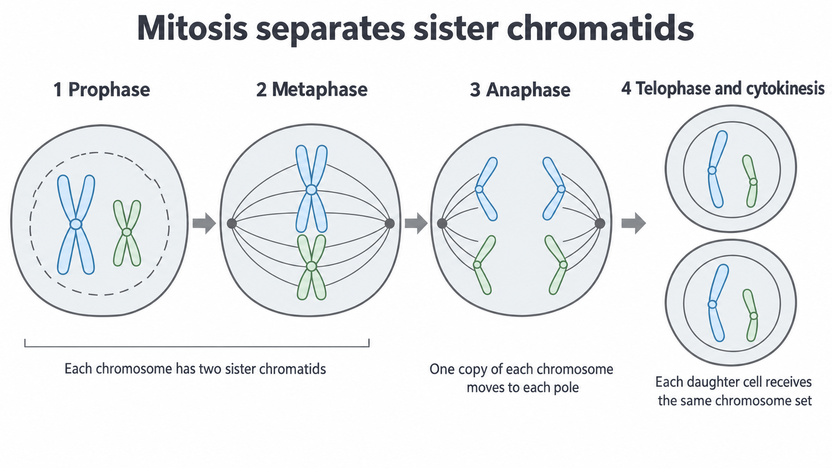
A simplified animal-cell sequence showing two representative chromosomes. The colours distinguish chromosomes, not homologous pairs. The final chromosomes remain drawn compactly so their allocation is visible; in real telophase they decondense. This is a schematic, not a micrograph.
The key event is the separation of sisters, rather than the separation of the two members of each homologous pair. Every duplicated chromosome contributes one copy to each daughter nucleus. This preserves the chromosome complement and normally preserves the alleles carried by the parent cell. DNA is not replicated again between metaphase and anaphase.
Cytokinesis then gives the two nuclei separate cytoplasms and cell-surface membranes. In an animal cell, a contractile ring draws the membrane inwards to form a cleavage furrow. In a plant cell, vesicles form a cell plate between the daughter nuclei; this develops into the new partition, including cell wall material. A rigid existing cell wall prevents a plant cell from simply pinching inwards like an animal cell.
Continue this lesson.
You’re previewing NovaLearn without an account. Create a free account to read the rest.
Create free account