8.8, 8.9 - Brain regions and medical imaging
A brain scan can show an abnormal structure, a change in metabolism or a response during a task, but these are different kinds of evidence. In 8.8 you will locate four major brain regions and connect them to their functions. In 8.9 you will use those connections to understand what MRI, fMRI, PET and CT can reveal in research and medical diagnosis.
Cerebral hemispheres and cerebellum
The large folded upper part of the brain is the cerebrum, divided into left and right cerebral hemispheres.
Cerebral cortex
The thin, folded outer layer of the cerebral hemispheres, containing many neurone cell bodies and involved in processing information.
Different cortical areas contribute to conscious sensory perception, voluntary movement, language, memory, learning and reasoning. For example, visual information reaches the visual cortex towards the back of the hemispheres, while motor areas send signals involved in voluntary movements. The folded surface provides a large cortical area within the skull. The hemispheres communicate through bundles of axons, including the corpus callosum; complex behaviour depends on connected networks rather than one isolated spot.
The cerebellum is a smaller, highly folded structure at the back of the brain, below the rear of the cerebral hemispheres and behind the brainstem. It coordinates the timing and precision of movements and helps maintain posture and balance. It uses sensory information about the body and movement-related signals to help adjust muscular activity. Initiating a voluntary movement and coordinating it are therefore different functions: a person may still attempt to reach for an object after cerebellar damage, but the reach may be poorly controlled.
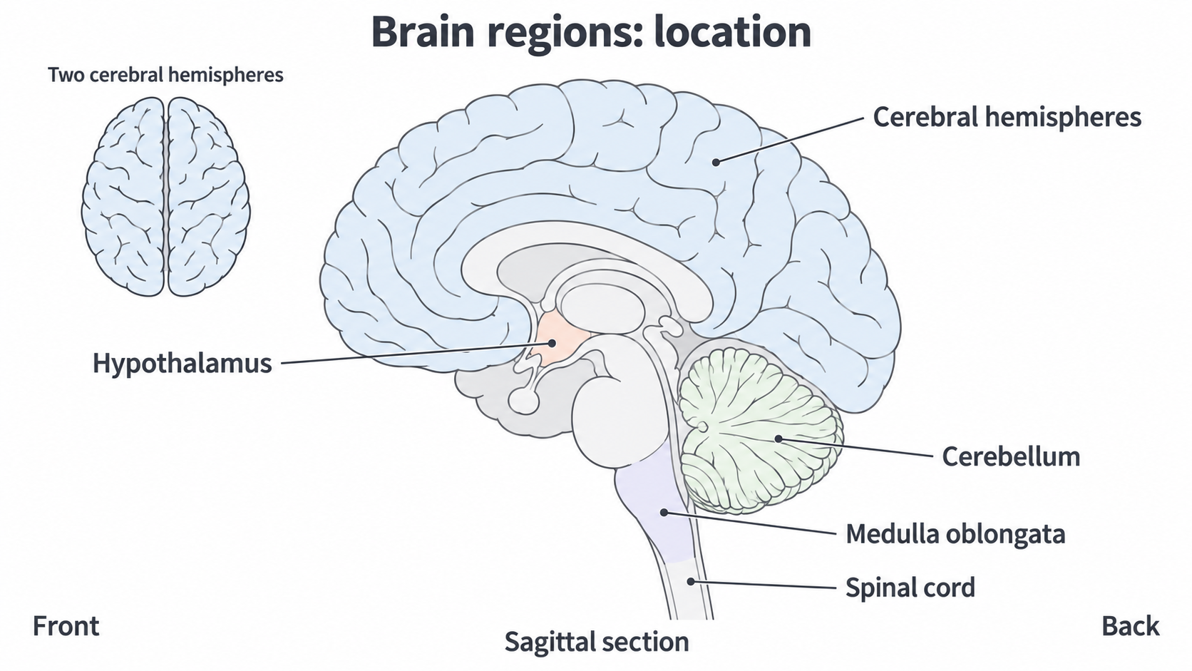
The sagittal view cuts from front to back through the midline. The inset shows why a side section alone does not display both hemispheres. This is a simplified anatomical illustration, not a patient scan.
Damage in a particular area can connect a structural observation with a functional difficulty. However, the difficulty depends on the precise tissue and connections affected; it is not enough to see a lesion somewhere in a large region and assume every function of that region is lost. The basic locations and functions are supported by NCI brain anatomy and NCI central nervous system anatomy.
Hypothalamus and medulla oblongata
Small deep regions of the brain help coordinate internal conditions and essential automatic activities.
The hypothalamus lies near the base of the forebrain, beneath the thalamus and above the pituitary gland. It helps maintain homeostasis, the regulation of internal conditions within suitable limits. It integrates information about internal conditions and coordinates nervous and hormonal responses, including control through the pituitary. Its functions include temperature regulation, hunger, thirst and the sleep–wake cycle. For example, when core temperature rises, hypothalamic control can promote sweating and increased skin blood flow, increasing heat loss. It connects the nervous and endocrine systems rather than operating as a separate hormone gland without neural input.
The medulla oblongata is the lowest part of the brainstem, below the pons, continuous with the spinal cord and in front of the cerebellum. It contains centres involved in automatic control of breathing, heart activity and blood pressure, and reflexes such as swallowing. These functions continue without conscious decisions. Its respiratory centres act within a wider network, including the pons: the medulla is not the only brain area that influences breathing.
The hypothalamus and medulla can influence overlapping body systems, but their roles should not be collapsed into one. The hypothalamus integrates homeostatic demands; medullary centres help produce and adjust vital automatic outputs. For example, changes in breathing and cardiovascular activity can occur without consciously deciding each breath or heartbeat. The heart's own pacemaker initiates its rhythm; brainstem signals modify heart activity rather than generate every beat.
Locate the hypothalamus deep near the brain's base above the pituitary; locate the medulla at the bottom of the brainstem where it joins the spinal cord. Link each position to its role, rather than using "controls the body" for both.
The location and homeostatic role are described in NCI central nervous system anatomy; MedlinePlus describes hypothalamic functions and Cleveland Clinic's medulla resource describes medullary automatic control.
Structural evidence from MRI and CT
Structural imaging reveals the position, shape and condition of brain tissue without opening the skull.
Magnetic resonance imaging (MRI) uses a strong magnetic field and radiofrequency pulses. Hydrogen nuclei, abundant in tissue water, respond to the field and pulses. As their magnetisation returns towards equilibrium, detectable radiofrequency signals vary with the tissue environment. A computer reconstructs detailed slices, which can also be combined into three-dimensional views. These differences allow good contrast between soft tissues, including grey and white matter.
MRI can help locate tumours, tissue damage or abnormal development and monitor changes over time. Ordinary structural MRI does not tell us what a person was thinking during the scan. It uses no ionising radiation, radiation energetic enough to remove electrons from atoms. This makes repeated structural studies possible without an x-ray dose. It still requires safety screening for implants and metal, can take longer than CT, is noisy and requires the person to remain still; motion can blur the result. NIBIB explains the signal, while ACR/RSNA describe brain MRI uses and limits.
Computed tomography (CT) uses x-rays sent through the head from many angles. Detectors measure the transmitted x-rays; different tissues attenuate, or reduce, the beam by different amounts. The computer reconstructs cross-sectional slices from these measurements. CT can rapidly show skull fractures and bleeding after a head injury, and can locate some masses or other structural abnormalities. It is useful when speed matters, but MRI generally gives better soft-tissue detail for many brain investigations. CT itself does not map the neurones active in a thought task.
CT exposes tissue to ionising x-rays, so the diagnostic benefit is weighed against a small radiation risk, especially for repeated exposure. This does not mean a necessary emergency CT should always be replaced by slower MRI. The medical question, speed, tissue detail and suitability for that patient all matter. These principles are described by NIBIB CT and ACR/RSNA head CT.
fMRI and the blood oxygenation signal
Functional imaging investigates changes associated with what the brain is doing, rather than only its visible anatomy.
Functional magnetic resonance imaging (fMRI) commonly measures the blood oxygenation level-dependent (BOLD) signal. Oxygenated and deoxygenated haemoglobin have different magnetic properties. Local neural activity can increase energy demand; blood flow normally increases in the active region, changing local blood oxygenation and the MR signal. The measured signal therefore depends on the blood response associated with activity. It is not a direct recording of action potentials, individual neurones or thoughts. The physical basis was demonstrated in Ogawa and colleagues' primary research.
Continue this lesson.
You’re previewing NovaLearn without an account. Create a free account to read the rest.
Create free account