4.9 - Starch, cellulose and microfibrils
A plant can build both a food reserve and a strong cell wall from glucose. This lesson explains how the form of glucose, the glycosidic bonds and the arrangement of chains make starch suited to storage and cellulose suited to support.
Glucose forms and glycosidic bonds
Starch and cellulose are both polymers of glucose, but the configuration of their glucose residues and the links between them differ.
Polysaccharide
A large carbohydrate made from many monosaccharide residues joined by glycosidic bonds. A residue is the part of a sugar molecule incorporated into the polymer.
α-glucose and β-glucose have the same molecular formula, , and the same connections between their atoms. They differ in the arrangement of the hydroxyl group, –OH, at carbon 1 (C1). In the glucose ring, C1 is the carbon next to the ring oxygen that also carries a hydroxyl group. C6 lies outside the ring in the –CH₂OH group attached to C5.
The reliable distinction is the position of the C1 hydroxyl relative to the C5-attached –CH₂OH group:
| Form of D-glucose | C1 hydroxyl relative to the C5-attached –CH₂OH group | In the usual Haworth drawing with –CH₂OH above the ring |
|---|---|---|
| α-glucose | Opposite face of the ring | C1 hydroxyl below the ring |
| β-glucose | Same face of the ring | C1 hydroxyl above the ring |
Rotating an entire drawing does not turn α-glucose into β-glucose: both groups rotate together, so their relative arrangement stays the same. In the source structures below, a solid wedge points towards the viewer and a hashed wedge points away. Compare the C1 hydroxyl at the upper right of each ring with the –CH₂OH side chain at the upper left. Unlabelled line ends and corners represent carbon atoms; most carbon-bound hydrogens are omitted.
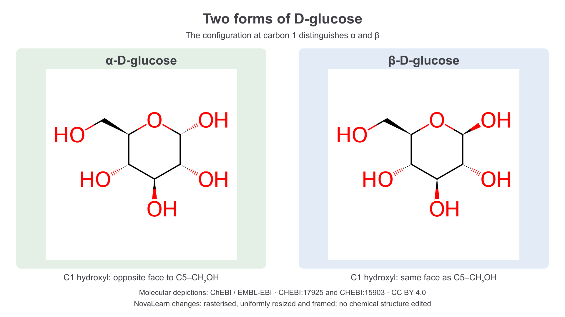
The two depictions differ at C1; the remaining stereochemistry is the same. Molecular depictions: ChEBI/EMBL-EBI, CHEBI:17925 and CHEBI:15903, CC BY 4.0. NovaLearn rasterised, uniformly resized and framed the complete depictions; no chemical structure was edited.
A glycosidic bond is a covalent linkage joining sugar residues through an oxygen atom. In the condensation model, an –OH group and an –H are removed from the reacting hydroxyl groups to form one water molecule as each linkage forms. Hydrolysis reverses this change: water is used to split a glycosidic linkage, restoring hydroxyl groups. Cells use enzyme-controlled pathways to build and break down these polymers; the condensation model describes their net chemical relationship rather than all the intermediate steps of synthesis.
The notation identifies the configuration and carbon positions involved. An α-1,4 linkage connects C1 of an α-configured glucose residue through oxygen to C4 of the next residue. A β-1,4 linkage connects the same numbered positions with the β configuration at the linked C1. A 1,6 linkage connects C1 to the external C6 position and can form a branch. Rotating a ring on the page changes neither its carbon numbering nor its linkage type.
Starch as a glucose reserve
Starch stores glucose-derived material in a compact form that a plant can mobilise when soluble sugars are needed.
Plant starch usually contains two α-glucose polymers: amylose and amylopectin. Their proportions vary between sources, so starch is not one molecule with a fixed composition.
| Component | Glycosidic linkages and arrangement | Consequence |
|---|---|---|
| Amylose | Long, largely unbranched α-1,4-linked chains | Chains can coil into helices, helping pack many residues into a small space |
| Amylopectin | α-1,4-linked chain segments with α-1,6 linkages at branch points | A branched molecule with many chain ends; segments can form helices and pack together |
The α-1,4 linkage gives a different chain conformation from the extended β-1,4 chain of cellulose. Starch chains associate into granules within plastids: chloroplasts can hold temporary starch reserves, while amyloplasts store starch in tissues such as a potato tuber. “Compact” concerns the packing of chains and granules; it does not mean each glucose residue has become smaller.
Starch is largely insoluble under normal cellular conditions. Storing glucose as an insoluble polymer therefore produces a much smaller osmotic effect than storing the same number of glucose units as free dissolved glucose. This helps a cell keep a large reserve without a correspondingly large increase in dissolved-particle concentration and water entry. Large insoluble granules are also retained in the cell rather than readily diffusing out through membranes.
When a reserve is needed, suitable enzymes break glycosidic bonds and release smaller soluble carbohydrates. Further processing can supply glucose for respiration or material for growth. The store is therefore mobilisable; “insoluble” does not mean “chemically impossible to break down”. For example, a germinating seed can use its starch reserve before its leaves can photosynthesise sufficiently.
Branching needs a careful explanation. Some starch-degrading enzymes work from chain ends. More branches provide more ends where such enzymes can act simultaneously, provided the ends are accessible and appropriate enzymes are present. However, α-1,6 branch linkages need suitable enzyme activity, and the packing and accessibility of the granule also influence breakdown. Branching alone cannot establish which of two real starch samples will be hydrolysed faster.
Starch combines compact packing, low solubility and controlled mobilisation. Explain each advantage through its effect on storage or sugar supply; do not treat branch number alone as a universal prediction of hydrolysis rate.
The storage advantage can be tested with a changed condition. If a cell replaced a starch reserve with the same number of free glucose molecules, there would be many more dissolved particles. This would increase the tendency for water to enter by osmosis, even though the total number of glucose-derived units was unchanged.
Cellulose chains and microfibrils
Cellulose forms long load-bearing structures in plant cell walls because its unbranched chains can lie closely alongside one another.
Continue this lesson.
You’re previewing NovaLearn without an account. Create a free account to read the rest.
Create free account