4.7, 4.8 - Plant cells and electron micrographs
A plant cell can make sugars, store starch and remain firm while surrounded by water, but different cell types share these jobs between them. In 4.7, you will connect plant-cell structures with their functions and compare them with animal cells. In 4.8, you will use genuine electron micrographs to recognise those structures and use their scale bars to judge size.
Plant and animal cell compartments
Plant and animal cells share the basic organisation of eukaryotic cells, but their outer boundaries and some internal compartments differ.
Both normally have a plasma membrane, cytoplasm, a nucleus containing chromosomes, mitochondria, ribosomes, endoplasmic reticulum and Golgi apparatus. Mitochondria carry out aerobic respiration in plant cells as well as animal cells. A green leaf still needs respiration: chloroplasts do not replace mitochondria.
Plant cells have a cell wall outside the plasma membrane. Many mature living plant cells also contain a large central vacuole bounded by a tonoplast. Plant cells can contain plastids, a group of organelles that includes chloroplasts and amyloplasts. Animal cells have neither cellulose cell walls nor plastids. Although animal cells contain small membrane-bound compartments, they do not normally have the large permanent central vacuole characteristic of a mature plant cell.
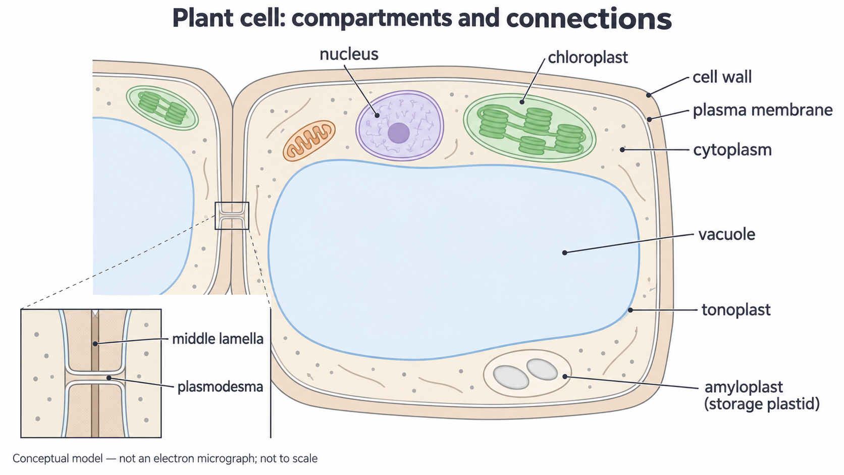
Conceptual model created for NovaLearn using ImageGen, 15 October 2026; not an observed cell and not to scale. It brings the named structures together for comparison. The adjacent-cell inset separates the two primary walls, the middle lamella between them and a membrane-lined plasmodesma connecting their cytoplasms.
The model is a map of possibilities, not a claim that every plant cell contains every organelle shown. A photosynthetic leaf cell has chloroplasts; an underground storage cell may have amyloplasts but no chloroplasts. Young dividing cells can have several small vacuoles. Some specialised mature cells lose structures during development, so absence of a nucleus in one section is not enough to classify a specimen as an animal cell.
Compare named structures and their evidence. A plant cell need not contain a chloroplast, and a plant cell containing chloroplasts still respires.
For example, a root storage cell and an animal liver cell may both have nuclei and mitochondria. A cellulose wall and starch-containing plastids support identification of the root cell; a rectangular outline alone is weaker evidence because sectioning and neighbouring cells affect shape.
Walls, middle lamella and pits
The cell wall provides mechanical support while the plasma membrane controls exchange with the living cytoplasm.
A primary cell wall contains cellulose microfibrils in a matrix of other polysaccharides. It is strong but can extend during growth. It resists the outward pressure produced when water enters the cell, helping prevent osmotic bursting and helping maintain shape. It is freely permeable to water and many small dissolved substances; it does not perform the selective transport role of the plasma membrane.
Middle lamella
A pectin-rich layer between the primary walls of adjacent plant cells that helps hold the cells together.
From one living cell to its neighbour, the sequence is cytoplasm → plasma membrane → its cell wall → middle lamella → the neighbour's wall → the neighbour's plasma membrane → its cytoplasm. These are different structures even when a low-resolution view merges several into one boundary. Some cells deposit a thick secondary cell wall inside the primary wall as growth ends. Such walls can be lignified; the detailed chemistry of cellulose and the organisation of supporting tissues are developed in the following lessons.
A pit is a local region where the secondary wall is absent or less thickened. Pits in neighbouring walls can face one another. The thin intervening primary-wall and middle-lamella material is called a pit membrane; despite its name, this is wall material, not a phospholipid plasma membrane. A pit therefore shortens the route through a thick wall. It is not necessarily an unrestricted hole through every cell boundary.
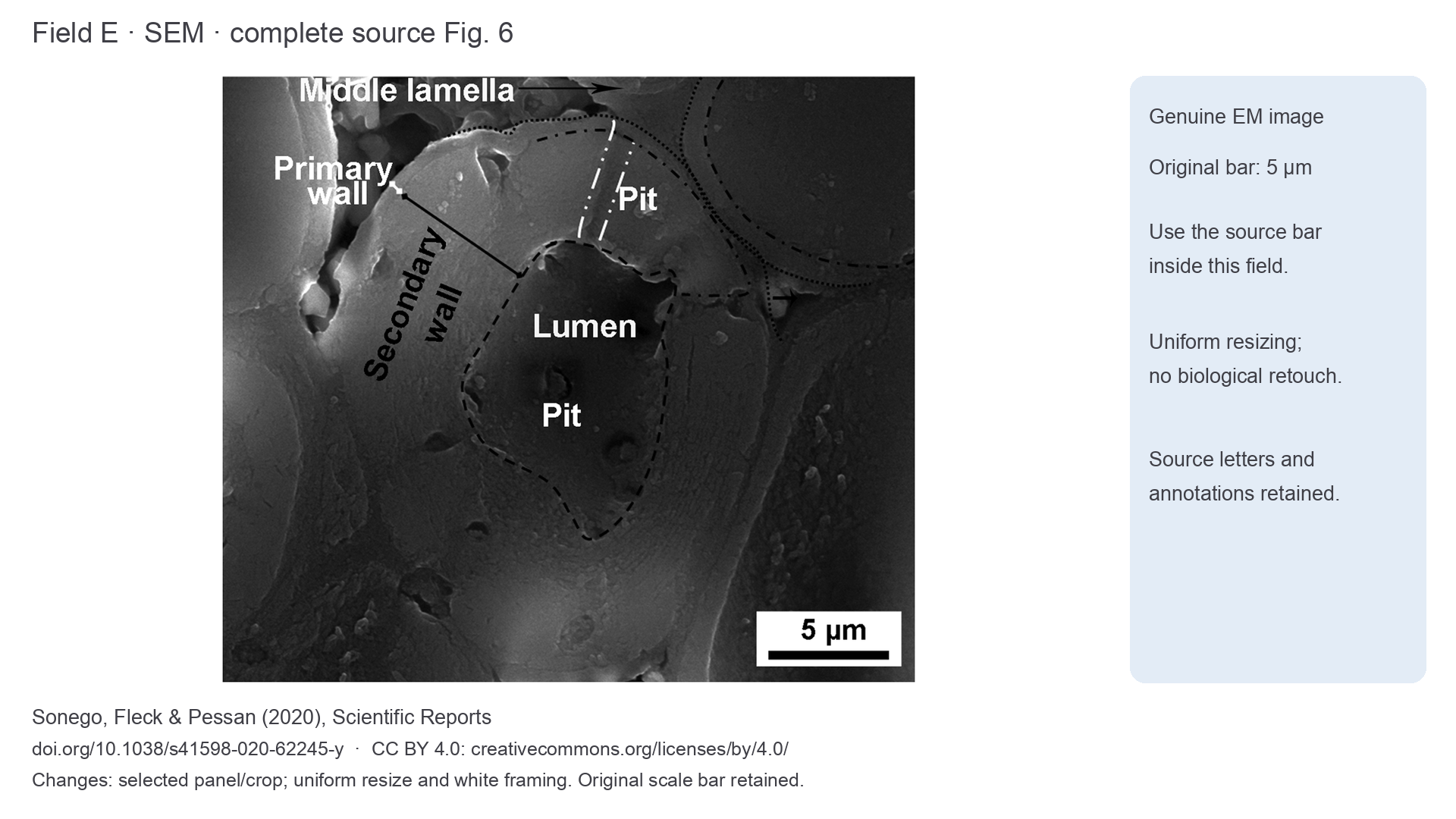
Field E: genuine SEM of a polished Brazil-nut mesocarp sclereid wall, Sonego, Fleck and Pessan (2020), Figure 6, CC BY 4.0. Changes: uniform resizing and white framing; the complete original image, source annotations and original 5 µm bar are retained.
In Field E, the lumen is the internal space within the thick-walled cell, not the wall itself. The broad secondary wall dominates the image. The source's marked pit crosses this thick layer; its outer end meets the thinner wall layers at the cell boundary. The middle lamella lies at the interface with the next cell, not inside the lumen. SEM shows surface relief; it does not reveal a living tonoplast simply because a dark space is visible. The composition and identity of these layers come from the researchers' wider evidence, not from greyness alone.
Plasmodesmata link cytoplasms
Plant cells can communicate directly through narrow channels across their shared walls.
Plasmodesma
A narrow, plasma-membrane-lined channel through a plant cell wall that connects the cytoplasm of adjacent living cells. The plural is plasmodesmata.
The plasma membrane is continuous through a plasmodesma. A narrow strand of ER commonly passes along its centre, leaving a cytoplasmic route around it. Small dissolved molecules and some signals can move from one cell's cytoplasm to the next; the channel's permeability is regulated. This helps cells coordinate activity while their walls hold them in place.
Pits and plasmodesmata are related but different. A pit is a region of reduced wall thickening. A plasmodesma is a much narrower living connection through the wall. Groups of plasmodesmata may occur in thin pit fields. A large depression in a wall is not, by itself, evidence that you can resolve an individual plasmodesma.
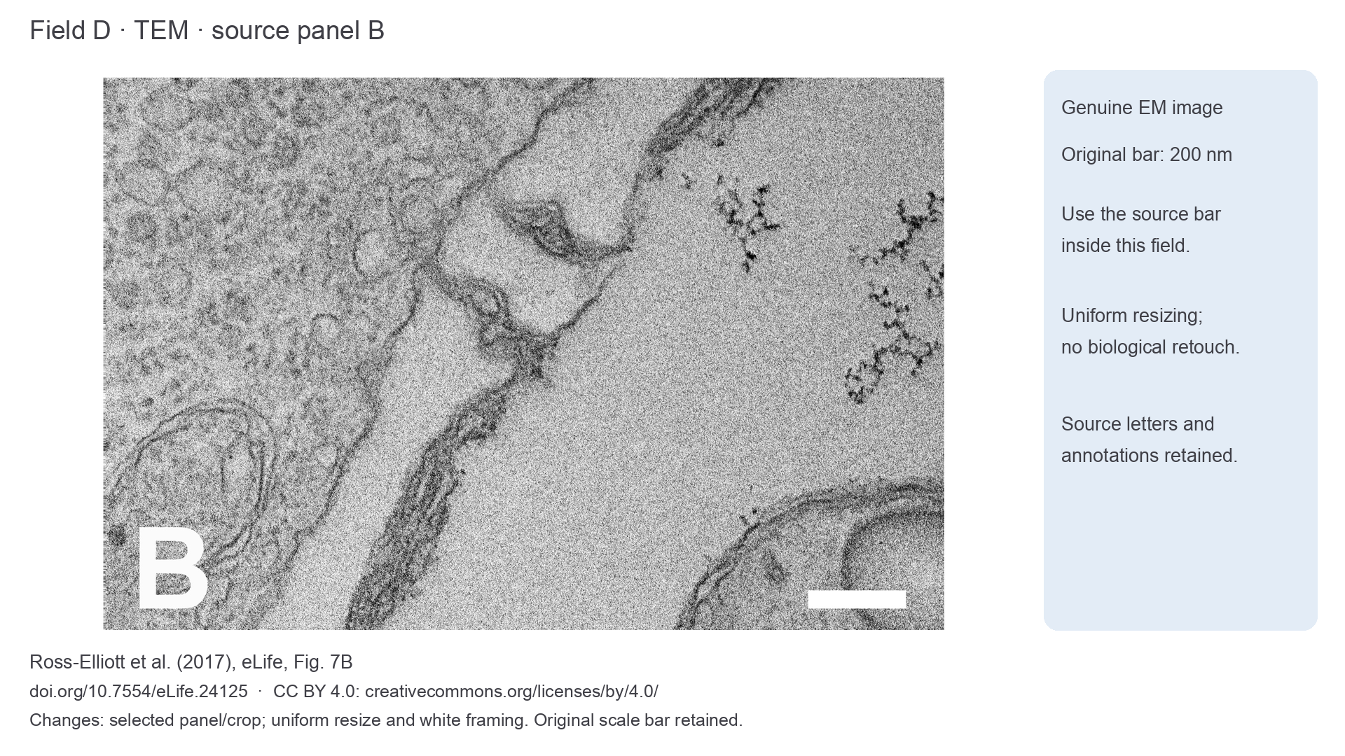
Field D: genuine TEM of a root-cell interface in Arabidopsis, Ross-Elliott et al. (2017), Figure 7B, CC BY 4.0. Changes: source panel B selected, uniformly resized and framed. The original 200 nm bar and source letter B remain; no specimen structures were erased.
Follow the broad wall running diagonally through Field D. Two much narrower bridges cross it rather than lying as free vesicles inside one cell. The wall on either side and continuity towards the two cytoplasms are the useful recognition features. The source identifies these as simple plasmodesmata. Their individual membrane components are harder to distinguish than the entire channel; do not invent a visible ER strand wherever the image cannot resolve one.
Animal cells can communicate through other types of cell junction, but they do not have plasmodesmata through cellulose walls. A comparison should name the specific structures rather than claim that only plants have communication between cells.
Chloroplast envelope and thylakoids
A chloroplast contains an organised membrane system that captures light and supports photosynthesis.
Its envelope consists of two membranes. Inside is the fluid stroma, containing enzymes, DNA and ribosomes. An internal membrane system forms flattened sacs called thylakoids; stacks are grana, connected by membranes between the stacks. Chlorophyll and other components for the light-dependent reactions lie in the thylakoid membranes. The large membrane area supports light capture and the reactions that transfer energy; enzymes in the stroma support sugar production. The reaction pathways are taught later in the course.
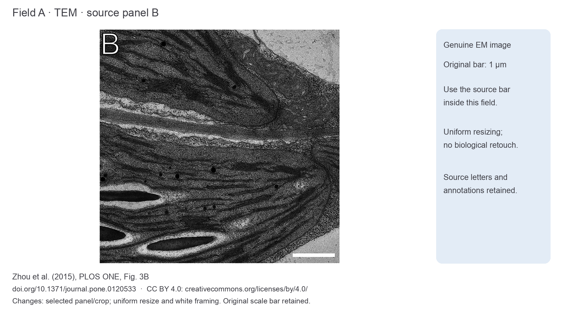
In a TEM, look for a bounded plastid with repeated closely spaced membrane profiles, often showing denser stacks within a lighter stroma. The section can make a stack look like parallel dark lines rather than a pile of discs. Chloroplasts are not green in ordinary TEM: brightness depends on preparation, staining and electron transmission, so colour cannot identify them.
Continue this lesson.
You’re previewing NovaLearn without an account. Create a free account to read the rest.
Create free account