2.9(iii), 2.9(iv) - Protein folding, haemoglobin and collagen
A polypeptide's amino acid sequence helps determine the shape it can form and the job it can do. In 2.9(iii), you will connect sequence, folding and the interactions that stabilise proteins. In 2.9(iv), haemoglobin and collagen show how different molecular structures suit oxygen transport and mechanical support.
Sequence and structural levels
A protein's structure can be described at different levels, from the order of its amino acids to the arrangement of several chains.
Primary structure
The specific sequence of amino acids in a polypeptide chain, joined by peptide bonds.
The peptide bonds form a continuous backbone. The variable R groups, or side chains, project from it. A sequence specifies which R groups occur and their positions along the chain. Some side chains are charged or polar; others are non-polar. Their size and chemical properties affect which regions of the chain can approach, interact and remain exposed to water. Two chains with the same length and the same numbers of each amino acid can therefore fold differently if their amino acids are in a different order.
Secondary structure is regular local folding of the backbone. An alpha helix is a coiled region of one chain. A beta pleated sheet contains extended regions lying alongside one another; these can be regions of the same chain folded back on itself or regions from different chains. Both are stabilised by hydrogen bonds between backbone C=O and N–H groups. These hydrogen bonds are different from the covalent peptide bonds that join adjacent amino acids.
Tertiary structure is the overall three-dimensional shape of one polypeptide chain. It includes its helices, sheets and less regular regions, brought into a particular arrangement. Interactions involving R groups help stabilise this shape. A compact folded chain is still one chain: folding has not made new polypeptides.
Some functional proteins contain more than one polypeptide. Each chain is a subunit. Quaternary structure describes how these subunits are arranged and interact. It is an additional level of organisation, rather than another name for the folding of a single chain. A functional protein made from one chain has no quaternary structure.
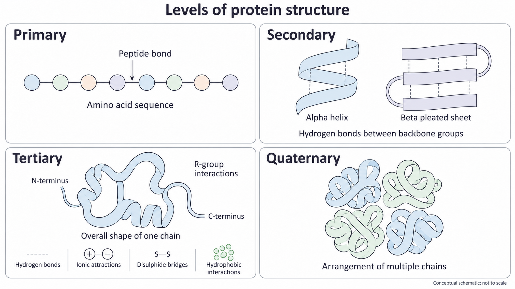
The four panels describe levels of organisation, not four separate proteins that must appear in every case. The interaction key lists possible contributors to tertiary structure; a particular protein need not contain all of them. This is a conceptual schematic, not an atomic model.
Interactions and folding
The folded shape depends on many interactions acting together, so changing an amino acid or the surrounding conditions can change a protein's properties.
The main interactions must be distinguished by what they connect and by their chemistry.
| Interaction | What it involves | Contribution to structure |
|---|---|---|
| Hydrogen bond | Attraction involving a slightly positive hydrogen attached to an electronegative atom and a nearby electronegative atom | Backbone hydrogen bonds stabilise helices and sheets; hydrogen bonds involving polar side chains also help stabilise the overall fold. |
| Ionic attraction | Attraction between oppositely charged R groups | Holds suitable regions of a chain, or different subunits, together. Changes in charge can weaken or remove it. |
| Disulphide bridge | A covalent S–S bond between the sulfur-containing side chains of two cysteine residues | Can link two regions of one chain or two different chains. It is a stronger covalent link, not a hydrogen bond or an ionic attraction. |
| Hydrophobic interactions | Non-polar R groups clustering away from surrounding water | Help bring non-polar regions together, often in the interior of a soluble, compact protein. This is not a new covalent bond between every pair of R groups. |
Weak attractions are individually less strong than covalent bonds, but many acting together can stabilise a folded protein. Close packing also allows weak van der Waals attractions between nearby atoms to contribute to stability. Peptide bonds remain the covalent links along the backbone. A disulphide bridge is a cross-link between side chains, so it does not replace the peptide bonds.
For example, suppose a positively charged side chain is close to a negatively charged one in a folded protein. Replacing the positive side chain with a non-polar one can remove that ionic attraction. The new side chain may also interact differently with water. This can alter the fold or the protein's surface properties. The effect depends on the position and nature of the change: a sequence change does not automatically destroy every protein's function.
Temperature and pH matter as well as sequence. Strong heating can disrupt interactions maintaining the fold. A large pH change can alter side-chain charges and therefore ionic attractions, and can affect hydrogen bonding. Denaturation is loss of the protein's normal three-dimensional structure, usually with loss of its normal function. Under ordinary denaturing conditions, the peptide backbone is not hydrolysed, so the primary sequence can remain intact. Breaking peptide bonds and cutting the chain is a different change. Loss of the normal fold can reposition binding groups or disrupt the contacts needed for mechanical support, explaining why function can be lost. Do not assume that heating breaks every disulphide bridge or that returning to the original conditions always restores function.
Sequence positions the interacting groups. Their interactions, in the surrounding conditions, stabilise the shape; the shape and surface chemistry help determine function.
This explains why knowing only the number of amino acids is insufficient to predict a protein's properties, and why an unchanged sequence can still lose its functional fold.
Globular and fibrous properties
Globular and fibrous proteins have different overall shapes and physical properties that suit different biological roles.
Globular proteins have compact, roughly rounded shapes. Many soluble globular proteins have hydrophilic, polar or charged groups on the outside, where they interact with water, and many hydrophobic groups buried inside. This arrangement helps the protein remain dispersed in an aqueous environment. Folding also brings particular groups together to make binding sites. Haemoglobin is a soluble globular protein inside red blood cells.
Fibrous proteins have elongated structures, often with repetitive sequences and arrangements. They commonly assemble into insoluble fibres that provide support or resist mechanical forces. Collagen forms such structures outside cells. A stable fibre can remain in place and transmit force through tissue rather than dissolve and disperse. These are useful general patterns, not a rule that every globular protein is soluble or every protein fits perfectly into one category.
Using observations to infer properties
The table gives original illustrative observations of two purified proteins under the same aqueous conditions.
| Observation | Protein P | Protein Q |
|---|---|---|
| Overall molecular shape | Compact and rounded | Long and narrow |
| Behaviour in water | Forms a solution of dispersed molecules | Remains in bundles |
| Surface or assembly observation | Polar groups exposed on the surface | Molecules overlap and are cross-linked |
P fits the usual soluble globular pattern: exposed polar groups can interact with water, supporting its dispersion. Q fits the fibrous pattern: overlapping, linked molecules can form a continuous load-bearing structure. The conclusions use both the observed shape and the physical behaviour. Shape alone would be weaker evidence for how the protein behaves in water.
These observations support a class of protein and a possible function. They do not establish that P is haemoglobin or Q is collagen. Identifying those specific proteins would need further molecular evidence, such as their sequences or characteristic structures. An insoluble bundle is not automatically proof of useful strength either: an incorrectly folded globular protein can aggregate.
Haemoglobin and oxygen transport
Haemoglobin combines a compact, soluble protein structure with specialised groups that bind oxygen reversibly.
The main adult human haemoglobin, haemoglobin A, has four polypeptide subunits: two alpha-globin and two beta-globin chains. Each has its own primary sequence and folded tertiary structure, with alpha-helical regions. The names alpha-globin and beta-globin identify two types of chain; a beta-globin chain is not a beta pleated sheet. Their association gives haemoglobin its quaternary structure.
Each folded subunit contains one haem group, a non-protein component held in a pocket in the globin. A haem group contains one iron ion, , which can bind one oxygen molecule, . Haem is a prosthetic group: a non-protein component closely associated with the protein and needed for its function. Haemoglobin is therefore a conjugated protein, containing both protein and non-protein components.
There are four haem groups in a complete haemoglobin A molecule, so it can carry up to four oxygen molecules, not just four oxygen atoms. The polypeptide sequence forms the appropriate pockets and subunit contacts; the haem iron provides the oxygen-binding site. A haem group is not an additional polypeptide subunit.
Reversible binding makes haemoglobin a transporter. Where oxygen availability is high, as in the lungs, oxygen binds. Where availability is lower, as in respiring tissues, oxygen can dissociate. Permanent binding would prevent delivery. Haemoglobin's hydrophilic outer surface helps it remain soluble in the aqueous cytoplasm of a red blood cell while its folded subunits hold the haem groups in suitable positions.
The subunits also influence one another. Oxygen binding changes haemoglobin's conformation and increases the affinity of the remaining sites for oxygen: cooperative binding. Interaction between subunits makes this possible. The molecule can therefore change its oxygen-binding behaviour without being denatured; a small functional conformation change is different from loss of its normal structure.
For a structure–function explanation, connect each feature to its consequence: four haem-containing subunits provide four binding sites; reversible binding allows loading and release; a soluble compact structure works in the aqueous environment inside red blood cells.
Collagen and tensile strength
Collagen's long molecules assemble into strong structures that resist pulling forces in connective tissues.
One collagen molecule contains three polypeptide chains wound around one another in a triple helix. Each chain adopts an extended helical conformation suited to this arrangement. It is not a standard alpha helix, and the triple helix is not three standard alpha helices wrapped together. Association of the three chains is organisation involving multiple polypeptides. Do not confuse a collagen molecule's three chains with the many collagen molecules in a fibril.
In the triple-helical region, glycine occurs at every third position in each chain. Glycine has a very small side chain, allowing close packing where the three chains meet. Many other positions contain proline or hydroxyproline, which help maintain the appropriate chain conformation and stability. Hydrogen bonds between chains help stabilise the triple helix. The primary sequence is therefore suited to forming a long, closely packed molecule rather than a compact globular one.
The chains do not have to be identical. For example, the common fibre-forming type I collagen has two alpha-1 chains and one alpha-2 chain. These are collagen-chain names; they do not mean standard alpha-helical secondary structure. Detailed synthesis and processing are not needed to explain how the mature fibre works.
Many collagen molecules lie alongside one another in overlapping, staggered arrangements to form fibrils. Covalent cross-links between molecules restrict them from sliding past one another; fibrils can be bundled into larger fibres. These structural cross-links must not be confused with hydrogen bonds within a triple helix or automatically called disulphide bridges. The strength arises from the assembled structure, with interactions at several levels, rather than from one exceptionally strong hydrogen bond.
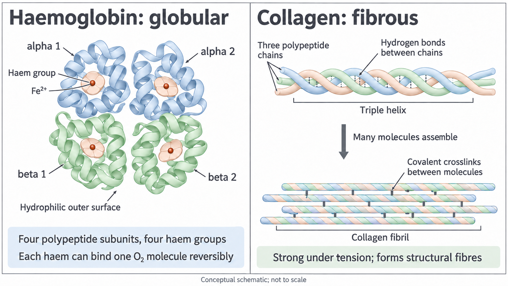
Haemoglobin's alpha and beta subunits are types of globin chain. The three colours in collagen distinguish individual chains, not necessarily three different chain types. Dashed links within a collagen molecule represent hydrogen bonds; solid links between molecules represent covalent cross-links. Both drawings are conceptual and not to scale.
Tensile strength means resistance to breaking under a pulling force. Collagen's long, overlapping, cross-linked assembly transfers load and resists stretching and rupture. In a tendon, this helps transmit the pulling force of muscle to bone. In ligaments and other connective tissues it helps maintain support. Collagen is insoluble as an assembled fibre, so it remains as a stable tissue component. It should not be described as an oxygen carrier or as a highly elastic protein that stretches and recoils like elastin.
Consider a change that leaves each triple helix intact but reduces the covalent cross-links between molecules. The chains within each molecule can still be held together, yet neighbouring molecules can slide more readily. The fibril may therefore resist a pulling force less effectively. This separates stability of one molecule from strength of the larger assembly.