8.10, 8.11 - Critical periods and visual development
Seeing depends on developing brain circuits as well as functioning eyes. In 8.10, you will learn how early visual activity refines connections; in 8.11, you will use Hubel and Wiesel's kitten and monkey evidence to judge what animal models reveal about human development.
Activity refines visual connections
Visual experience changes the effectiveness and organisation of connections in the developing visual cortex, the region of the cerebral cortex that first processes much of the information arriving from the eyes.
Critical period
A developmental interval when a particular circuit or capacity is especially sensitive to experience, so that abnormal or missing experience can have lasting effects.
Light produces changes in retinal signalling. Information travels through visual pathways, including a relay in the thalamus, to the primary visual cortex. Connections within and beyond this cortex help extract features of a scene. Vision therefore requires a working pathway and appropriate processing, rather than an eye simply transmitting a finished picture.
Synaptogenesis
The formation of synapses between neurones.
Developing circuits initially form many connections. Some are subsequently retained and others eliminated, a process called synaptic pruning. Measurements of synapses in developing monkey cortex provide evidence for initial overproduction relative to adult levels. Pruning a synapse does not necessarily kill the neurone that formed it. Rakic and colleagues' primary study
The initial organisation is not a blank slate waiting for the first image: inherited developmental processes and activity within the nervous system establish a starting circuit. Experience then helps refine it. When an input repeatedly contributes to activity in a target neurone, its synapses can become more effective and stable. Less effective inputs may weaken and lose connections. This is an activity-dependent process, rather than a conscious decision by the brain to keep useful pictures. Research on coincident inputs from the two eyes supports a role for the timing of activity in this refinement. Primary research on binocular input activity
With suitable patterned input from both eyes, connections supporting both eyes can be maintained. When one eye supplies much less useful patterned input, its influence can weaken relative to the other eye's influence.
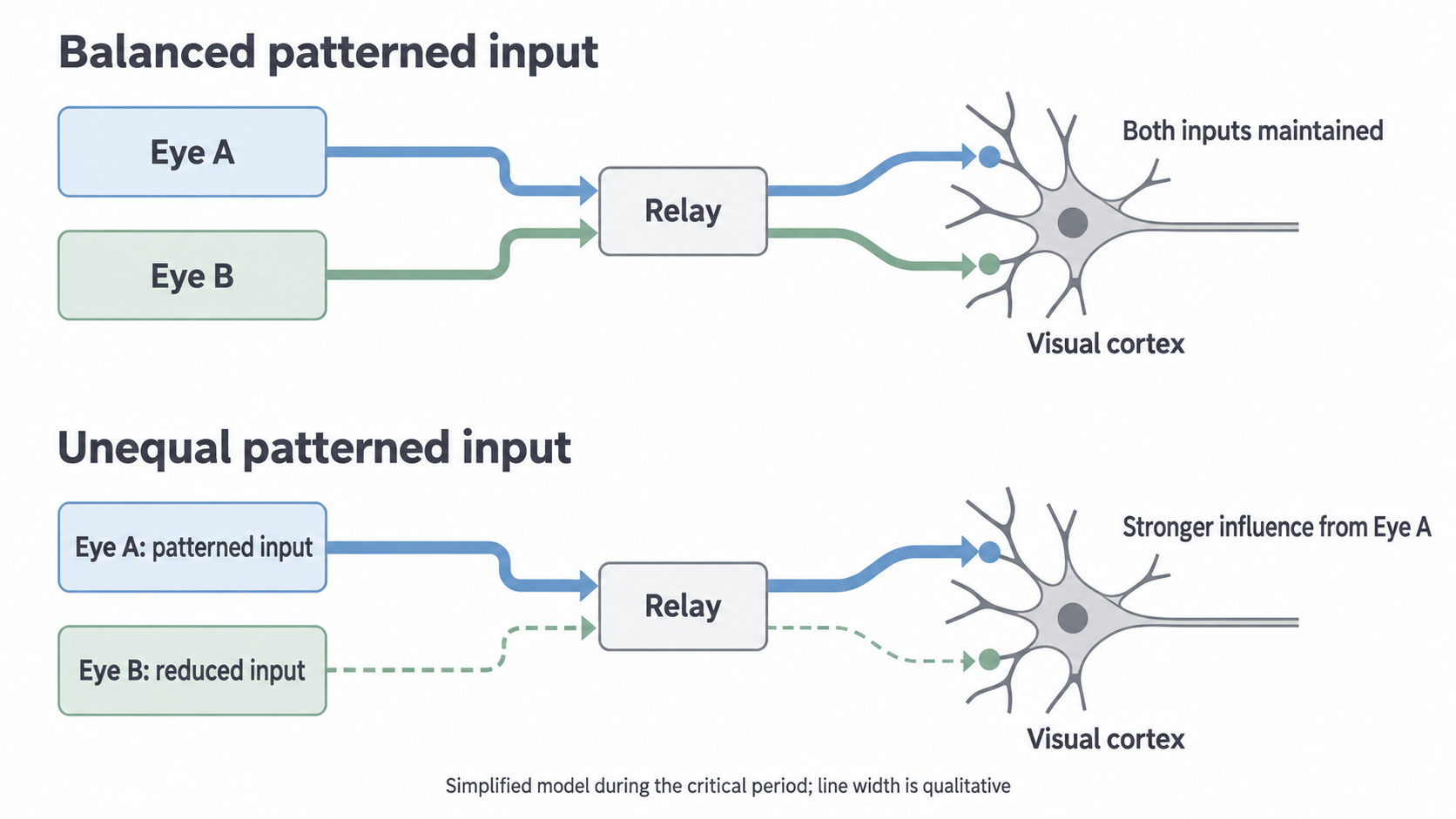
A conceptual circuit during the critical period. The relay represents intervening visual pathways, not a direct retina-to-cortex connection. Line widths show relative influence qualitatively; this is neither a measured tracing nor a complete anatomical map.
A window of heightened plasticity
The same disruption of input can have different consequences at different developmental ages because the capacity for circuit change itself develops.
Plasticity
The capacity of nervous-system connections and function to change.
During the visual critical period, patterned visual activity helps refine synaptic strengths and the distribution of inputs. Normal experience through both eyes supports the full development of visual capacities, including the coordinated use of both eyes. Binocular vision uses information from both eyes; comparison of their slightly different views contributes to depth perception.
Ocular dominance describes the relative influence of the two eyes on a cortical neurone's response. Many neurones in normally developed binocular regions respond to either eye, although not necessarily equally. Early unequal input can shift this balance towards the eye receiving useful patterned stimulation, leaving fewer neurones effectively influenced by the other eye.
The critical period involves both opportunities for refinement and vulnerability to disruption. It is not simply the interval in which the greatest number of synapses exists. Maturation of connections and inhibitory circuits helps change plasticity with age. For example, primary mouse research links maturation of initially silent excitatory synapses to the ending of a period of ocular-dominance plasticity; the molecular detail is supporting evidence, rather than a list to memorise. Primary study of synapse maturation
Timing, duration and the function measured all matter. A brief disruption at high susceptibility may have a larger effect than a longer disruption later. Reopening a pathway does not instantly rebuild a previously altered circuit. Conversely, “critical period” does not mean all later learning or recovery is impossible. Different properties can have different sensitive windows, and ages measured in one species are not a calendar for another.
Full visual development requires appropriate activity while circuits are especially plastic. An intact eye alone does not guarantee that its cortical connections develop normally.
What the kitten studies measured
Hubel and Wiesel used animal models to test whether early visual input affects cortical development, rather than merely observing that vision improves with age.
In Wiesel and Hubel's 1963 study, seven kittens and one adult cat experienced monocular deprivation: one eye was prevented from receiving normal patterned vision. Deprivation lasted between one and four months, with starting ages varied. The investigators subsequently compared cortical single-cell responses when each eye was stimulated. The deprived eye influenced far fewer cortical cells than expected from normal comparisons; the other eye dominated. This was evidence of altered cortical processing, not simply a test of whether a closed eyelid blocked light at that moment. These are historical observations, not an investigation to reproduce. Original 1963 paper
Later experiments varied onset age and duration more deliberately. The following is an original evidence summary, not a new dataset or a complete inventory of animals.
| Historical comparison | Reported cortical finding | What the comparison contributes |
|---|---|---|
| Deprivation during the fourth and fifth weeks of kitten life for only three or four days | A marked loss of binocularly driven cells and a shift towards the non-deprived eye | A short exposure can matter during high susceptibility. |
| Similar disruption initiated at later ages | Susceptibility declined after the early peak | Developmental age changes the effect. |
| An adult cat deprived for more than a year | No detectable comparable physiological effect | Duration alone cannot explain the juvenile effect. |
These findings support a sensitive early interval, rather than a simple rule that more days without normal input always produces a larger change. Hubel and Wiesel's 1970 report also found limited cortical recovery after prolonged early deprivation even when the eye was reopened. The reported age boundaries refer to their particular physiological measurements; they should not be treated as universal limits for every aspect of cat vision. Original age-and-duration study
Continue this lesson.
You’re previewing NovaLearn without an account. Create a free account to read the rest.
Create free account