3.3.13.2 - Proteins
Proteins are built from amino acids, but the interesting part is what happens next: those amino acids join into chains, fold into specific shapes, and can be broken back down and analysed. This lesson keeps those ideas connected so that peptide formation, protein structure, hydrolysis, and chromatography all feel like parts of one story.
From amino acids to peptide links
Each amino acid contains both an amino group and a carboxylic acid group. That means one amino acid can react with another: the carboxyl group of one joins to the amino group of the next, and a molecule of water is removed. This is a condensation reaction, and the new link formed between the two residues is what turns separate amino acids into a peptide chain.
Peptide link
A peptide link is the link formed when the carboxyl group of one amino acid reacts with the amino group of another amino acid.
In proteins, amino acids are really just repeated units in a chain. Two amino acids make a dipeptide. Three make a tripeptide. A long chain is a polypeptide, and proteins are made from one or more long polypeptide chains. The sequence matters: glycine-alanine is not the same as alanine-glycine, because the order of residues in the chain is different.
To draw a peptide, use this method every time:
- Write the amino acids in the correct order.
- Remove from the carboxyl group of one amino acid and H from the amino group of the next.
- Join the carbonyl carbon to the nitrogen to make .
- Keep the amino group at one end of the chain and the carboxylic acid group at the other.
For example, glycine and alanine form the dipeptide:
A tripeptide contains two peptide links. In general, a chain made from amino acids contains peptide links, which is a useful quick check when you sketch or count bonds in a chain. The diagram below shows how glycine, alanine, and serine join by condensation to form a tripeptide with two peptide links.
[DIAGRAM: asset_name: 3.13.2 - Proteins - Diagram 1; asset_slug: 3.13.2 - Proteins - Diagram 1; recommended_method: retained_png; description: Condensation formation of a tripeptide from glycine (H2N-CH2-COOH), alanine (H2N-CH(CH3)-COOH), and serine (H2N-CH(CH2OH)-COOH). Show the three amino acids before reaction, highlight removal of OH and H at each join, then show the final tripeptide H2N-CH2-CONH-CH(CH3)-CONH-CH(CH2OH)-COOH with both -CONH- peptide links clearly labelled and 2H2O formed.]
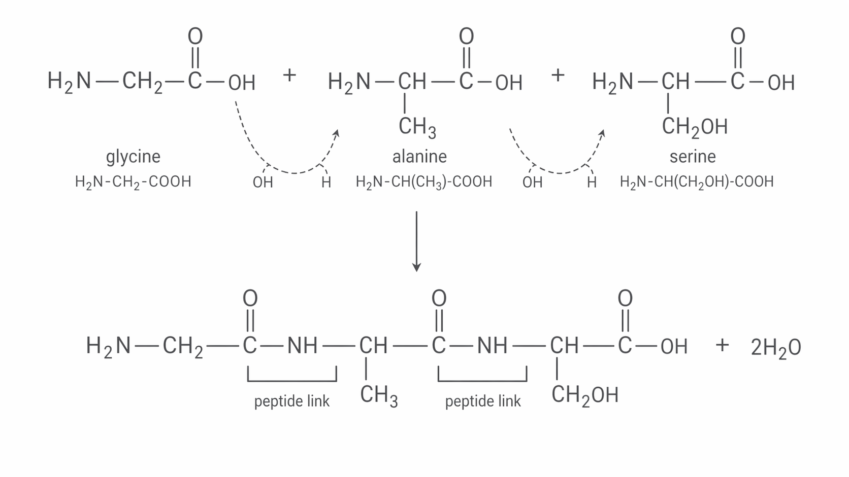
One common mistake is to draw the chain and forget that the end groups stay different. The left-hand end still has an amino group, and the right-hand end still has a carboxylic acid group. That is why a peptide can keep reacting to form a longer chain.
For the order alanine–glycine–serine, keep the side chains on their original alpha carbons:
There are two C(=O)–NH links and two water molecules are eliminated. Reversing the order gives a different structure even though the molecular formula is unchanged. The serine side-chain OH is retained when drawing the specified peptide backbone.
Hydrolysis breaks peptide links
Hydrolysis is the reverse of condensation. Water is used to break the peptide link, so the peptide or protein is split back into its constituent amino acids. If every peptide link in the chain is hydrolysed, the final mixture contains the amino acids that originally made up the protein.
Hydrolysis
Hydrolysis is a reaction in which a bond is broken by reaction with water.
For peptide hydrolysis, imagine cutting the chain at each link. The from water ends up on the carbonyl side, reforming a carboxylic acid group, and the H ends up on the nitrogen side, reforming an amino group. If a tripeptide is hydrolysed completely, three amino acid molecules are produced.
A reliable way to draw hydrolysis products is:
- Identify each amino acid residue in the chain.
- Break every bond in a peptide link.
- Add to the carbonyl carbon side and H to the nitrogen side.
- Rewrite the resulting amino acids as separate molecules.
So if the peptide is hydrolysed, the products are glycine, alanine, and glycine. The key skill is reading each residue correctly before you split the chain.
In the laboratory, complete hydrolysis is often carried out by heating with hydrochloric acid. That detail matters because strongly acidic conditions can protonate amino groups in the mixture. For understanding the structure, though, the central idea is simply that hydrolysis regenerates the amino acids that formed the peptide in the first place.
Primary and secondary structure
Once you move from a short peptide to a protein, there is more than one level of structure to think about. You need to distinguish clearly between the order of amino acids in the chain and the shapes that the chain adopts.
The primary structure is just the sequence of amino acids along the polypeptide chain. If even one amino acid is in a different position, the primary structure has changed. Primary structure is held together by covalent peptide links, not by hydrogen bonds. That distinction is important, because students often mix up what holds the chain together with what makes it fold.
Secondary structure
Secondary structure is the regular arrangement of a polypeptide chain into an alpha-helix or beta-pleated sheet, held in place by hydrogen bonds.
In an alpha-helix, the chain coils into a spiral. In a beta-pleated sheet, sections of the chain lie next to each other in a folded sheet. In both cases, the stabilising hydrogen bonds form between and groups in the backbone of the polypeptide. The amino acid side chains are not the main point here; the repeated backbone is what makes these regular patterns possible. The diagram below compares the primary structure with the two regular forms of secondary structure.
[DIAGRAM: asset_name: 3.13.2 - Proteins - Diagram 2; asset_slug: 3.13.2 - Proteins - Diagram 2; recommended_method: code_generated; description: Primary residue sequence, schematic alpha coil and beta sheet, plus explicit carbonyl O lone-pair dotted attraction to H–N.]
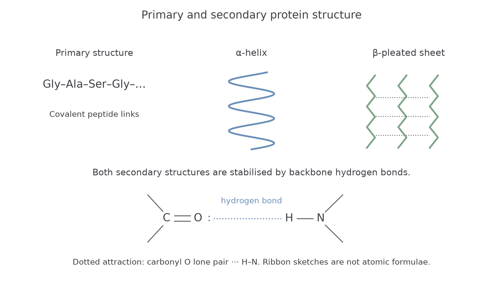
When you identify structures in diagrams, look for the visual clue that matches the level. A simple sequence of residues or amino-acid abbreviations suggests primary structure. A spiral suggests alpha-helix. A folded sheet with repeated strands suggests beta-pleated sheet. Both the helix and the sheet are examples of secondary structure.
In a detailed drawing, the hydrogen bond runs from the carbonyl oxygen to a hydrogen on N–H: . The oxygen supplies a lone pair; the N–H hydrogen is partially positive. Do not connect C to H, O to O, or N directly to the carbonyl carbon with a dotted “hydrogen bond”. The covalent backbone remains intact when these weaker attractions are disrupted.
Continue this lesson.
You’re previewing NovaLearn without an account. Create a free account to read the rest.
Create free account