6.5.3 - Ultrasound imaging, impedance and Doppler
Ultrasound imaging turns reflected sound pulses into information about position, tissue boundaries and blood flow. In this lesson you will connect the wave physics to the scanner: how a transducer sends and receives pulses, why boundaries reflect some ultrasound, why gel matters, and how the Doppler shift can be used to measure blood speed.
Ultrasound and transducers
Ultrasound is sound, so in body tissue it travels as a longitudinal mechanical wave: particles of the medium oscillate parallel to the direction of energy transfer. The word ultrasound means the frequency is above the upper limit of human hearing.
Ultrasound
Ultrasound is a longitudinal wave with frequency greater than .
Medical scanners use a probe called a transducer. The active part is a piezoelectric crystal. When an alternating potential difference is applied across opposite faces of the crystal, it expands and contracts rapidly, so it emits ultrasound pulses. The same crystal can also act in reverse: when an echo compresses or stretches it, a small potential difference is produced and detected by the electronics.
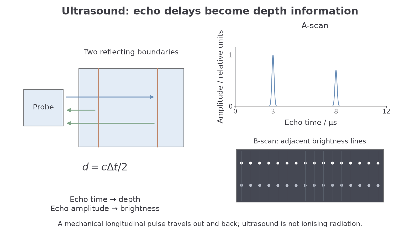
That dual behaviour is why one probe can both send and receive. A short pulse is sent into the body, then the probe waits for echoes from boundaries between media. The time delay tells the scanner how deep the reflecting boundary is, and the echo amplitude gives information about how strong the reflection was.
Pulse-echo A-scans and B-scans
An ultrasound pulse travels to a boundary and then the echo travels back to the transducer. If the pulse has speed in the tissue and the echo from a boundary returns after time , the depth of that boundary is:
Pulse-echo distance
The factor of 2 is essential: the measured time is for the out-and-back journey.
An A-scan is the simplest display. It shows echo amplitude against time. Each spike represents a reflected pulse; a later spike is from a deeper boundary, and a taller spike is from a stronger reflection. OCR questions often use A-scans for measurements such as eye-lens thickness, where two echoes come from the front and back surfaces of the lens.
A B-scan builds an image from many pulse-echo measurements. The probe either sweeps across the body or uses many small transducer elements. For each scan line, echo time fixes depth and echo amplitude fixes brightness. Combining many brightness lines gives a two-dimensional section through the tissue.
[DIAGRAM: asset_name: 6.5.3 - Ultrasound imaging, impedance and Doppler - diagram 1; asset_slug: 6_05_3_ultrasound_scanning_impedance_and_doppler__diagram_01; recommended_method: drawn_physics; description: Two wall depths match 3 and 8 μs A-scan peaks; first stronger echo appears brighter in B-scan; echo arrows return from the relevant walls.; caption: Echo time determines boundary depth, while echo amplitude determines brightness in a B-scan image.]
Worked example: an A-scan of an eye lens shows two echoes from the front and back of the lens. The time difference between the echoes is . Take the speed of ultrasound in the lens as .
The ultrasound travels across the lens and back between those two echoes, so:
Convert the time difference:
Substitute:
So the lens thickness is about .
A-scans are useful for precise distances between identified boundaries, such as the front and back of an eye lens. B-scans show a cross-section, useful for viewing soft tissue and fetal development in real time. Ultrasound does not use ionising photons, and a portable scanner can be comparatively inexpensive. However, air and bone can block useful transmission; a strong surface reflection can hide deeper structures. Higher frequency can resolve finer detail but is usually attenuated more strongly, limiting useful depth. The method must fit the required depth and anatomical information.
Acoustic impedance and reflection
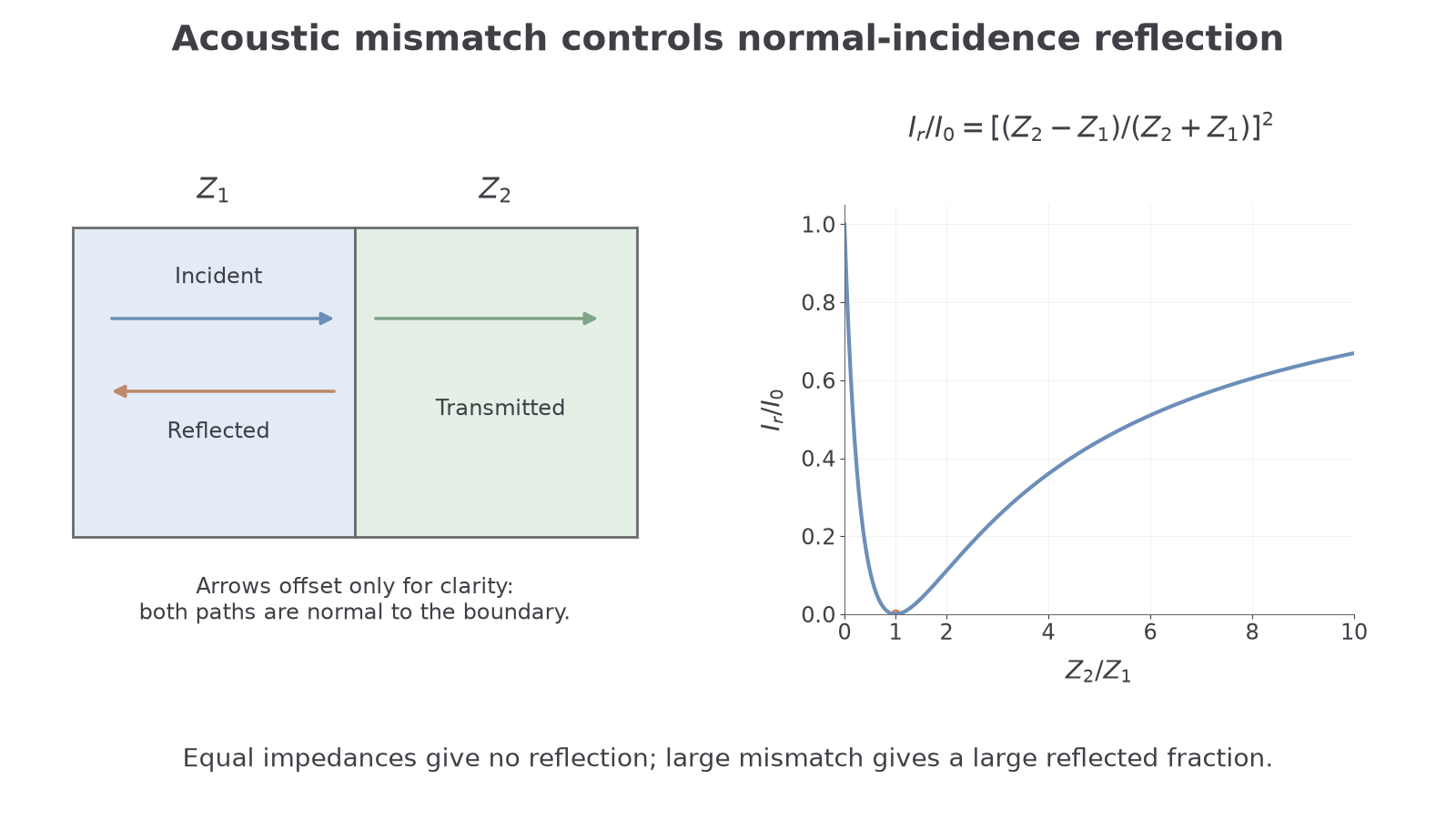
When ultrasound reaches a boundary between two media, such as two different tissues, some intensity is reflected and the rest is transmitted. The reflection depends on the acoustic impedance of the media.
Acoustic Impedance
The acoustic impedance of a medium is the product of its density and the speed of ultrasound in that medium.
Acoustic impedance
The unit follows directly from the defining equation:
If the acoustic impedances on the two sides of a boundary are and , the fraction of incident intensity reflected at normal incidence is:
Intensity reflection coefficient
Here is the incident intensity and is the reflected intensity. This ratio has no unit. If , the numerator is zero, so there is no reflection at that boundary. If the difference between the impedances is large, a larger fraction is reflected.
[DIAGRAM: asset_name: 6.5.3 - Ultrasound imaging, impedance and Doppler - diagram 2; asset_slug: 6_05_3_ultrasound_scanning_impedance_and_doppler__diagram_02; recommended_method: drawn_physics; description: Incident/reflected paths normal to interface and offset only for visibility; analytic mismatch curve has R=0 at Z2/Z1=1.; caption: At normal incidence, reflected intensity depends on the acoustic-impedance mismatch. Equal impedances give no reflection.]
Continue this lesson.
You’re previewing NovaLearn without an account. Create a free account to read the rest.
Create free account