6.5.1(e)-(g) - X-ray imaging and CAT scans
Plain X-ray images and CAT scans both use X-rays, but they answer different medical questions. In this lesson you will see how contrast media such as barium and iodine make selected structures stand out, how a CAT scanner builds slice images using a rotating X-ray beam and detectors, and why OCR expects a more precise comparison than just "CAT gives a clearer image".
Contrast from attenuation
The useful idea from X-ray attenuation is simple: different materials remove different fractions of the incident X-ray beam before it reaches the detector. Bone attenuates X-rays more strongly than soft tissue, so fewer photons reach the detector behind bone. On a common medical display, strongly attenuating regions are shown as pale or white, while regions that transmit more X-rays are shown darker. Some digital displays can invert the grey scale, so the physics statement is safer than memorising "white" or "black": greater attenuation means a different detector signal.
X-ray image contrast
X-ray image contrast is the visible difference between regions in an image caused by different transmitted X-ray intensities reaching the detector.
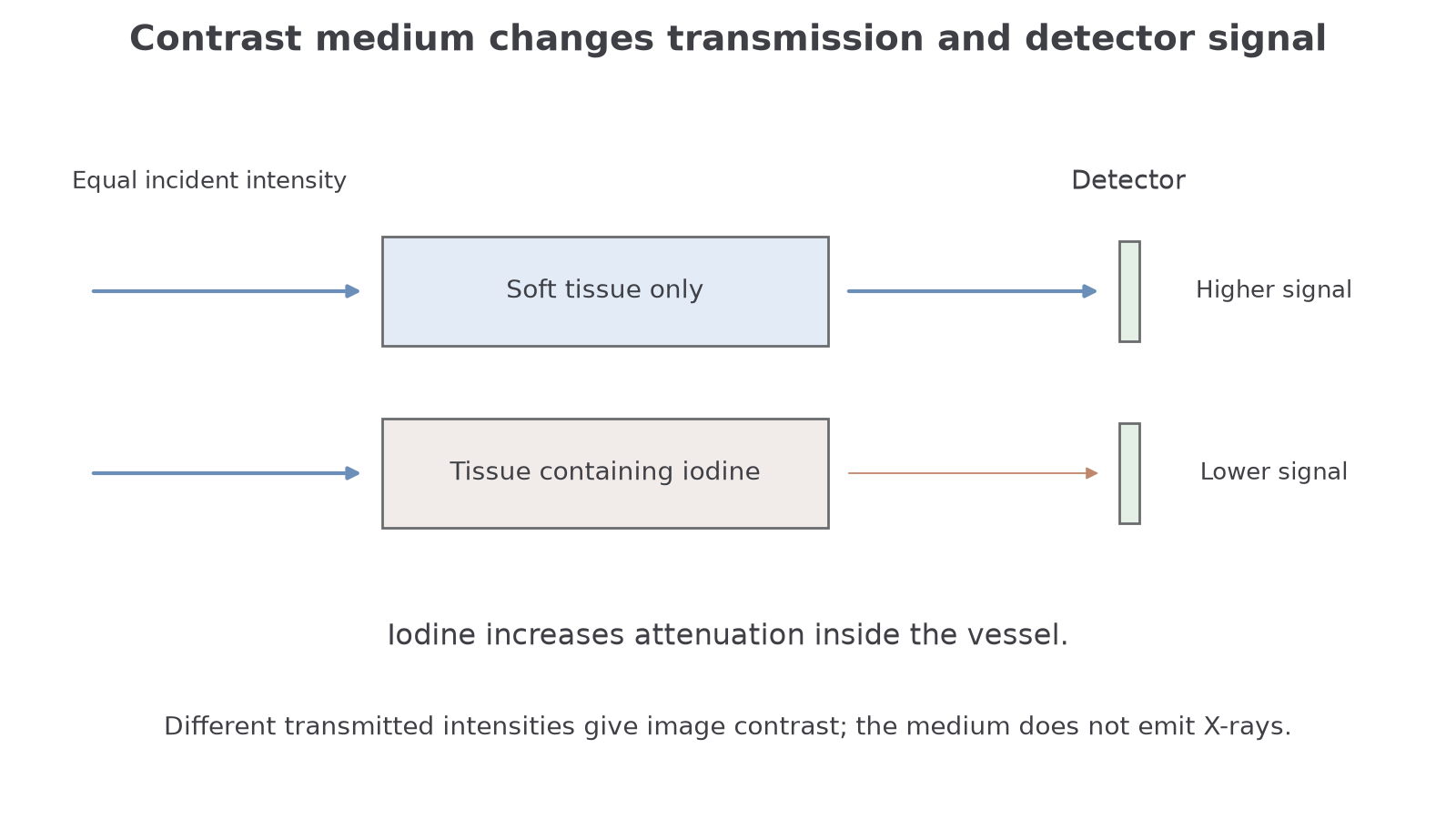
Soft tissues often have similar attenuation coefficients, so their detector signals can be too similar to distinguish clearly. A contrast medium is a substance introduced into a particular part of the body to change the attenuation there. It makes the target structure have a different transmitted intensity from neighbouring tissue.
Barium and iodine are useful because atoms of high proton number interact more strongly with X-rays than the low-proton-number atoms common in soft tissue. The contrast medium does not make the body radioactive; it changes how strongly part of the body attenuates the X-ray beam.
The image is still a projection image: every point on the detector receives the result of X-rays passing through a whole line of tissue. This is why contrast media are powerful but not magic. They can make one structure stand out, but structures along the same beam path may still be superimposed.
[DIAGRAM: asset_name: 6.5.1(e)-(g) - X-ray imaging and CAT scans - diagram 1; asset_slug: 6_05_1b_xray_imaging_contrast_media_and_cat_scans__diagram_01; recommended_method: drawn_physics; description: Equal incident beams traverse equal path lengths; iodine-containing material gives smaller transmitted detector signal.; caption: Iodine increases attenuation, reducing the transmitted detector signal compared with surrounding soft tissue.]
Barium and iodine
Barium and iodine are common X-ray contrast media. Their high-proton-number atoms attenuate X-rays strongly, and suitable compounds can be introduced into the region being investigated.
Contrast medium
A contrast medium is a substance introduced into the body to increase image contrast by changing how strongly selected tissues or spaces attenuate the imaging radiation.
Barium is usually used as a barium sulfate compound for the digestive system. It can be swallowed to outline the oesophagus, stomach or intestines, or given rectally for the lower bowel. The important physics is that the barium-containing region has much greater X-ray attenuation than the soft tissue around it, so the shape of the digestive tract can be inferred from the detector pattern.
Iodine-based contrast media are often used for blood vessels and soft-tissue organs. They may be injected into a vein or artery so that the contrast medium is carried by the blood, or used in other routes depending on the clinical investigation. Again, the physics is contrast by attenuation: the iodine-containing region changes the transmitted X-ray intensity.
Two distinctions explain their imaging role.
First, "contrast" does not mean the medium gives out radiation. The X-ray source provides the ionising radiation. The contrast medium changes the absorption pattern.
Second, barium and iodine are not interchangeable labels to sprinkle into any answer. Barium is the standard example for the gut; iodine is the standard example for blood vessels and organs where a soluble contrast agent is needed.
Contrast media bring HSW benefit-risk reasoning into the topic. They can make a diagnosis possible without surgery, but patients are screened for suitability because adverse reactions can occur and some iodine-based agents need care for people with kidney problems or previous contrast reactions. The A-level point is not to learn clinical protocols; it is to recognise that useful diagnostic evidence must be balanced against risk.
CAT scanner components
CAT means computerised axial tomography; CT means computed tomography. The scanner combines many measurements with a reconstruction algorithm.
CAT scan
A CAT scan is an X-ray imaging technique in which X-ray measurements from many angles are processed by computer software to produce cross-sectional slice images, which may be combined to form 3D information.
The assessable components are:
- a rotating X-ray tube;
- a thin fan-shaped X-ray beam;
- a ring of detectors;
- computer software;
- a display.
The thin fan-shaped beam passes through a slice of the patient. The detector ring measures the transmitted X-ray intensity for many paths through that slice. As the X-ray tube rotates around the patient, the computer receives measurements from many angles. Software reconstructs a cross-sectional image from those measurements and sends the result to the display.
Continue this lesson.
You’re previewing NovaLearn without an account. Create a free account to read the rest.
Create free account